Causes. Consequences. Treatment.
There are several reasons for the appearance of calluses on a dog's elbows.
In this matter, a lot depends on the nutrition that the animal receives! A lack of animal fats in the diet and a lack of fat-soluble vitamins (A, E, D) lead to the skin on the dog’s elbows becoming rough, cracking, and calluses appearing. Calluses also quite often appear due to hard bedding or when the dog sleeps on a bare floor, stone, or wooden surface.
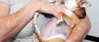
If your dog is overweight, sooner or later he will develop calluses on his elbows, and the heavier the dog, the higher the risk of developing calluses.
There is some breed predisposition - for example, Labradors, NO, VEO, KO and SAO suffer from this more often than other breeds. The sexual cause is more weakly expressed, however, in large males this problem occurs 2-3 times more often than in females, this is due to the presence of a female sex hormone that affects the elasticity, nutrition and hydration of the skin. For the same reason - the lack of female sex hormones, in sterilized bitches whose ovaries have been removed, this problem occurs more often than in unsterilized ones, but there is no clear statistical data on this issue.
Diet against calluses on the elbows in dogs.
There is only one conclusion from all that has been said: it is necessary to provide your dog with good, high-quality, balanced nutrition, rich in fat-soluble vitamins, control its weight, and monitor where your dog sleeps and lies. It’s good if you add one teaspoon or tablespoon (depending on the dog’s weight) of homemade unrefined vegetable oil to your dog’s food every day. Olive or linseed oil, as well as a mixture of them, are perfect. Vegetable oil will restore liver function and improve the condition of the dog’s skin. You can also order high-quality salmon oil, for example Salmon Oil, or give children purified fish oil without additives in the autumn-winter period.
The diet of such dogs must include red fish (a soup set of salmon, the back and head of pink salmon, a soup set of trout), cottage cheese should be taken from 3.5-9% inclusive, twice a week give 1-2 raw chicken yolks or 2 raw quail eggs with shell, also once a week, beef liver is fed, raw pink salmon heads, freed from gills, help well.
Liquid vitamins A and E in a bottle can be added to food in a course for 14 days, the calculation is based on weight, as for children. It is advisable to take a biochemistry test first, because fat-soluble vitamins are dangerous due to their overdose and can accumulate in tissues, and their excess, like excess oils, leads to malfunctions of the liver and pancreas. Therefore, such advice is not suitable for chronically ill animals, with pancreatic problems and gastrointestinal diseases. You can use the usual drug “Aevit” in capsules for this purpose; it should be given by weight 1-2 capsules X 2 times a day, for a course of 14 days with meals, if there is a sufficient amount of liquid.
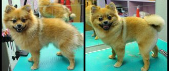
Often the problem is that owners do not attach importance to this “disease” and when calluses appear in their dog, they do not take any action, considering this almost the norm or a breed feature. Although it is during this period of time, at the initial stage, that it is enough to take minimal measures to solve or significantly improve the condition of the dog’s elbows - reduce weight, provide the correct covering for the animal to rest, establish nutrition and supply vitamins A, E for 14 days, elbows can simply Lubricate with olive oil daily for a month.
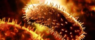
In the future, this problem worsens, places of ulceration appear, sometimes exudate and ichor leak from micro-wounds, swelling and pain appear, this creates a dangerous opportunity for the addition of a secondary infection (bacterial or fungal), or a mixed etiology. Therefore, if there is a chronic course, it makes sense to take a scraping for the fungus.
Another important reason could be high blood sugar levels and diabetes. Eliminate all sweets and fruits with high sugar content from your diet, and take a clinical blood test. This analysis and check of blood sugar levels in recently neutered males is mandatory; indirect signs are drooping eyes and sudden weight gain.
Option 1.
If calluses are roughened, keratinized skin that often cracks and sometimes suppurates, the following should be done. We clean the surface, calluses on the elbows are treated twice a day with a broad-spectrum antiseptic (not peroxide), this can be: “chlorhexidine”, “miramistin”, “betadine”, “povidone-iodine”, “octenisept”. Then, an antibacterial ointment (“levomekol”, “levosin”, “malavit” or “dimexide”) is applied to the naturally dried surface. Using ointments containing antibiotics for more than two weeks is not advisable!
Option 2.
If you see only cracks and slight swelling without suppuration, instead of antibiotic ointment, use an emollient and moisturizing cream or use wound healing creams: “bepanten-ratiopharm”, “panthenol”, etc. If the surface is very wet, instead of ointments, you should use Baneocin powder; it is better to leave it without covering, but do not allow the dog to lick it off, using a protective collar at home if necessary.
I do not recommend using Vaseline; it forms a surface film, which prevents the skin from breathing and creates a favorable environment for the development of anaerobic bacteria.
Option 3.
In more severe cases, when healing occurs slowly, Clotrimazole or Triderm ointments are added to the treatment, applying them in a thin layer. They also practice “elbow masks” twice a week. For this purpose, take a regular baby cream and add a small amount of Vit A, E, add 1 teaspoon of sea buckthorn oil, put a sterile 16X14 napkin (or a 7x7 self-adhesive “Cosmopor” napkin) on top, secure the edges with a fixing plaster, preferably Peha-haft " If the medicine has two forms of release, you should choose a thicker ointment rather than a cream!
The tips listed above also help with scuffs and cracks in the pads on dogs’ paws.
The healing process varies and may take several months. It is also important not to confuse a true callus with one formed after an injury; it is characterized by noticeable swelling, pain, sometimes this place can be hot, it is necessary to find the site of injury, measure the animal’s temperature and it is advisable to consult a doctor. There are also inflammatory diseases in the area of the joint capsule - bursitis or inflammation of the tendons - purulent tenosynovitis, then fluid also accumulates.
when using materials
News edited by: maugli — 6-03-2020, 12:50
Mostly, calluses develop in the elbow and hock joints (back legs). These calluses are dry, scaly, hairless and hard. Some are dark brown and black, and some are grey; it depends on the dog's skin and how long he has had the callus. Just like in humans, dog calluses are caused by the skin being in contact with something hard for an extended period of time, over time it becomes rough, cracked and sometimes requires veterinary attention. What are calluses on the elbows in dogs and what treatment is applicable to them can be read below.
Healing of fractures
In the first days after a fracture, no recovery processes or, conversely, resorption processes determined by radiography are established and bone fragments on the radiograph appear the same as they were formed during the injury.
Then, if no complications occur, around the ends of the fragments on the side of the periosteum, as a result of the proliferative activity of cells, a primary connective tissue callus is formed, connecting the distal and proximal bone fragments into one whole. This callus is not visible on the image because it absorbs X-rays to the same extent as the surrounding soft tissue.
The connective tissue callus gradually ossifies and gives thick, delicate shadows on the radiograph. The edges of the fragments at this time are somewhat decalcified and the intensity of the shadow approaches that of young bone tissue. During this phase of callus formation, the fracture gap remains clearly visible on the image and even expands. This depends on the partial resorption of the damaged part of the bone substance at the ends of the fragments.
Subsequently, the young bone tissue and decalcified edges of the fragments become increasingly ossified, their shadows become denser and clearer, merging into a single uniform dense tissue - callus. This callus undergoes complete ossification within 4-6 weeks. However, it is necessary to take into account age, location of the fracture in the bone, the degree of displacement of fragments and disruption of the periosteum, method of treatment, etc.
Otherwise, healing of a fracture occurs in the presence of many fragments lying around the fracture site. Bone fragments that remain connected to the periosteum are involved in the formation of callus. (Bone fragments that do not maintain sufficient connection with healthy tissues become necrotic and are rejected in the form of sequestra.
Dead bone fragments, or sequestra, are identified on an x-ray by the following features: greater density of their shadows compared to surrounding living fragments, absence of signs of new bone formation along their periphery, the presence of granulation tissue separating sequestra from living tissue.
Such complications, on the one hand, slow down the healing of the fracture, on the other hand, constant irritation of the periosteum caused by infection leads to the formation of a voluminous callus. But the most dangerous complications during fracture healing are osteomyelitis and the formation of a pseudarthrosis.
Traumatic osteomyelitis is a purulent inflammation of the bone marrow involving all elements of the bone and surrounding soft tissues with the formation of necrosis.
Osteomyelitis caused by trauma is radiographically characterized by the appearance of sharp bone destruction of a focal or even widespread nature, and the formation of sequesters in the cavities.
Osteomyelitis is usually accompanied by the formation of fistulas. To study them, the fistulography method is used. A contrast agent is injected into the fistula canal, after which X-ray photographs are taken. Sterile aqueous paste of barium sulfate is used as a contrast agent. To study fistulas associated with serous cavities, iodolipol is used. The contrast agent is injected into the external opening of the fistula using a syringe. Images of the subject being examined body parts are made in two mutually perpendicular projections.
In the case of a favorable osteomyeletic process, bone restoration proceeds according to the type of healing of bone defects. However, the bone does not always return to its previous shape; deformations often remain in the form of swelling, curvature, sclerosis, and periosteal layers.
False joint (pseudoarthrosis) is persistent abnormal mobility at the site of a former fracture, resulting from a disruption in the process of callus formation (M. V. Plakhotin).
The reasons for the formation of a false joint are: significant loss of bone substance along with the periosteum, incorrect position of bone fragments (bone fragments are adjacent to each other with their lateral surfaces or are far apart), lack of timely and correct immobilization of fragments, muscle fibers getting into the space between the fragments, long-term purulent processes and a number of other general and local causes.
The main radiological symptoms of a pseudarthrosis are: absence of callus between fragments, rounding and grinding of the ends of fragments, closure of the medullary canal.
Clinical symptomatology of acute forms of purulent arthritis makes it possible to make an accurate clinical diagnosis - purulent arthritis.
While the purulent inflammatory process is localized in the synovial membrane of the joint capsule, it is not possible to detect any abnormalities in the articular ends of the bones and cartilage on the x-ray, except for the expansion of the x-ray joint space. Only when articular cartilage and articular surfaces of bones are involved in the process do X-ray studies acquire their practical significance. At this time, the image shows a narrowing of the joint space as a result of the destruction of cartilage and the convergence of the bones that form the joint. Sometimes bone sequestra separated from the articular surfaces are visible in the joint cavity. Local defects, irregularities and patterns on the periarticular edges of the bone shadows are clearly visible. Subsequently, if effective treatment measures have not been taken, purulent inflammation begins in the bones of the joint. In this case, the disease is called purulent osteoarthritis. X-rays at this time reveal an increase in the joint space, osteolysis and foci of destruction, and at the same time increased bone growths are noted.
source
Types and consequences of calluses
Some pet owners consider dog calluses to be inherently pathological, but experts have different opinions. In some cases, the hardened tissue becomes additional protection for the elbow tendons. If calluses are left untreated, they crack and bleed, resulting in lesions (pain). If a pressure ulcer is left untreated, it is more likely to become infected. If calluses are oozing or bleeding, it is best to schedule an appointment with your veterinarian to rule out any problems.
Calluses are usually not a serious problem, but they can develop into ulnar hygromas. Initially, a callus on the elbow may form to protect the elbow from injury, but excessive and repeated stress on the joint can lead to a fluid-filled growth called an ulnar hygroma. With any luck, the hygroma will go away on its own within a few weeks if the dog has soft bedding and a small area. However, if the growth becomes infected, the veterinarian will need to drain the exudate from it.
Callus dermatitis is also common. Most often, they overcome dogs of large breeds, since in this case the pressure on the elbows is much stronger, but individuals with short legs sometimes suffer from the disease. If your dog has short hair, he is even more susceptible to the disease. This type of dermatitis in dogs differs from ordinary calluses in that when the surface of the callus cracks, an infection enters the microcracks, which, without timely intervention, can severely damage the skin. Purulent fistulas, boils, and ulcers may form. This will cause a lot of inconvenience for the dog, plus it can lead to blood poisoning or an abscess.
Therapy for callus dermatitis is also aimed at softening and healing, but requires the help of antibiotics. This type of disease is completely cured no earlier than after 2-3 months, and it is necessary to constantly show the dog to a doctor. The doctor will examine the pathological material to understand how the therapy is progressing. At home, you can use ointments against bacteria, which your doctor will recommend. Self-medication in such a situation can only delay the process and allow the infection to spread.
What is a callus and what does it look like after a fracture?
The process of bone tissue restoration is quite complex and multi-stage. The regenerative abilities of bones help restore lost functions after a fracture. Before the bone restores its integrity, a special multicellular formation is formed - callus. It fixes bone fragments and creates conditions for rapid growth and reproduction of bone cells. The article discusses how this formation is formed, why it is necessary, and what to do if the process goes wrong.
Therapy
Preventing calluses from forming on your dog's elbows is a long process. Lying on soft bedding or carpets can help prevent blisters, but some dogs prefer the cool tiles of any expensive orthopedic bed, especially during the warmer months. During the colder months, calluses often disappear because dogs prefer to lie on warmer, softer bedding.
Luckily, there are many medications for elbow calluses that can help. Today, oil is one of the most effective remedies. You can take almost any known oil or make a mixture of several. Good for therapy:
- sunflower;
- olive;
- apricot;
- linseed oil.
Vegetable oil can additionally be mixed into your dog’s food.
The oil is rubbed into the affected area several times a day for one month. Even if you rub the oil twice a day, the result will be visible after a week. This method is completely safe, because natural products are used for treatment, which can be absorbed by the dog when licked.
Therapy using Vaseline is another proven method. Vaseline will quickly soften the affected area. Apply Vaseline generously to your dog's calluses and rub in thoroughly. Massaging and rubbing the product into calluses is an important point. It is necessary to penetrate into the structure of the callus, and not just touch its surface. Apply every 12 hours for a week, and then the affected area is additionally treated with Vaseline to soften and completely heal.
Using Vaseline can leave an unpleasant aftertaste because the dog's elbows will stain the furniture at home, leaving greasy marks. Therefore, it is better to cover the lubricated area with a bandage. The low price and effectiveness of the product will help you set priorities.
Coconut oil is a good remedy for softening and healing calluses. It is necessary to rub it into the affected area for 2-3 minutes. The downside may be that the dog will want to lick such a delicious-smelling product. Again, a bandage will help. Vitamin E has a similar effect and is widely used for home therapy. These products also soften calluses in animals well.
No matter what product you use, start moisturizing your dog's elbow calluses as early as possible for faster results.
Reasons and what could it be?
Have you been trying to heal your JOINTS for many years?
Head of the Institute for the Treatment of Joints: “You will be amazed at how easy it is to cure your joints by taking the product for 147 rubles every day.
Professional dog breeders have repeatedly wondered why their beloved pets develop unpleasant calluses? One of the main reasons is a hard sleeping surface. Typically, the ulna bones are in constant contact with the ground. While resting or following commands, the dog comes into close contact with the hard surface of the ground or floor, thereby thinning the delicate skin. This problem is common among large dogs and, in particular, among males. This is due to the fact that their body contains fewer female hormones responsible for the elasticity and firmness of the skin. Therefore, the larger the animal, the greater the likelihood of calluses forming.
Our readers successfully use Sustalaif to treat joints. Seeing how popular this product is, we decided to bring it to your attention. Read more here...
As dog breeders have noticed, over time, areas in the elbow area begin to go bald, then growths appear in the form of hardened skin with darkening and roughness.
In fact, some calluses may be the primary symptoms of a dangerous disease such as callus dermatitis.
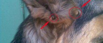
When examining a dog, special attention should be paid to the appearance of the ulnar plaques. The presence of bloody ulcers, ulcers, swelling or tissue compactions may indicate that this is a more serious pathology. In this case, the pet must be immediately shown to the veterinarian, especially if touching the growths causes discomfort or pain to the animal.
Another cause of calluses in dogs is poor nutrition and lack of vitamins A, E, D in the body. As a rule, a pet's diet should be rich in healthy fats, proteins and carbohydrates, the absence of which can lead to roughening of the skin, hair loss, and the formation of growths in the area of the elbow bones. You should not ignore the primary manifestations of calluses, since in the early stages of the development of the disease you can achieve complete disappearance of the problem.
The danger of calluses for an animal
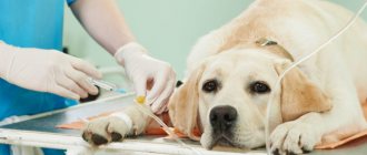
Many owners do not consult a doctor when they discover calluses, considering them a problem only from a cosmetic point of view. But calluses must be treated. When walking, they can cause pain and discomfort to the animal. The dog often licks wounds on the elbow joints and there is a risk of pathogenic bacteria and organisms getting into it, which will lead to inflammation and the development of callus dermatitis. In addition to rough, gray skin, if untreated, ulcers appear, accompanied by itching and pain.
Skin diseases in dogs
Based on its condition, the owner can determine whether the pet is healthy or sick. The following factors have a negative effect on the skin:
- Hormonal imbalance.
- Hypovitaminosis.
- Cardiac pathologies.
- Unbalanced diet.
The following types of skin diseases are distinguished:
- parasitic;
- fungal;
- bacterial;
- allergic.
Most often, diseases such as demodicosis and sarcoptic mange are detected in animals. These pathologies are characterized by severe course and long-term treatment.
Demodectic mange is dangerous because the mite spreads through the lymph system and spleen.
Often the mite is encapsulated in the roots of the fur. To destroy it, the animal is shorn. This provides medications with access to all layers of the skin. With timely treatment, you can get rid of parasites in 2–3 weeks.
To clarify the diagnosis, the veterinarian first takes a scraping from the affected area. Most often, ticks live on:
- body;
- head;
- paws.
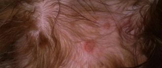
The main symptom of sarcoptic mange is active hair loss. The skin darkens, folds form on it, and dandruff appears. The affected areas are very itchy, which leads to scratching.
Sarcoptic mange is treated with medication. In some cases, the disease is transmitted to the owner. Parts of the body that come into contact with the dog begin to itch painfully.
Microsporia is usually diagnosed in dogs. This pathology is caused by the fungus Microsporum canis. Identifying the disease is quite simple. The veterinarian places the dog under an ultraviolet lamp. The fungus living on the skin is highlighted in a greenish color. If the need arises, the sick animal is sent for laboratory examination.
Many fungal pathologies are dangerous to humans.
After clarifying the diagnosis, the veterinarian prescribes drug therapy. Taking medications is combined with baths. And products are also used for local treatment of affected areas of the skin.
Long-haired dogs are cut. In order to prevent relapse, the room in which the pet lives is thoroughly disinfected. Bedding and toys need to be replaced.
The most common bacterial pathology is pyoderma.
If the disease is not treated, the infection spreads to:
- head;
- muzzle;
- lips;
- spaces between teeth.
Symptoms may occur in puppies. You need to sound the alarm when specific black dots appear.
The disease is long and difficult to treat. The dog is prescribed antibiotics. Drug therapy is accompanied by bathing and treating the skin with antiseptic drugs.
Neglected bacterial pathologies contribute to the development of abscesses. Anemia may result.
Allergy refers to the reaction of antibodies that are present in a dog’s body to irritants. The result of this is histamine activity. Reacting with blood particles, it provokes the development of the inflammatory process.
An allergic reaction is caused by:
- medicines;
- food;
- fleas;
- allergens.
The most dangerous medications are considered to be the penicillin group.
Often the allergic reaction is immediate. It can also appear 2-3 hours or several days after contact with the irritant.
Psoriasis on the elbows - symptoms
Psoriasis is characterized by the appearance of inflammatory rashes of varying diameters on the elbows, tending to grow peripherally. In the initial stages, these can be single elements, which, in the absence of proper treatment, merge, forming large psoriatic plaques and extensive diffuse lesions. On the surface of the inflammation, silvery scales are clearly visible, easily removed by mechanical action. When scraping, along with peeling, a thin film is removed (the film terminal phenomenon) and a pink-red surface is exposed, on which drops of blood appear (the blood dew phenomenon). In some patients, psoriasis is accompanied by the appearance of papules, around which a rim devoid of scales forms. The disease has a wave-like nature, in which periods of remission are replaced by periods of exacerbation. Regression is characterized by a weakening of the color intensity of the inflammatory elements until complete disappearance. Resorption of plaques, as a rule, begins from the central part, and in their place depigmented areas are formed. During the period of incomplete remission, individual lesions of small diameter remain on the elbows. In advanced stages, large diffuse lesions are not subject to significant changes for a long time. This is the so-called chronic form of psoriasis, which is very difficult to treat. Sometimes the disease is complicated by arthritis (arthropathic psoriasis), which is characterized by severe swelling and damage to the joints, damage to the nails (formation of pinpoint pits in the form of a thimble).
In most patients diagnosed with psoriasis, their general health does not deteriorate, and the disease is relatively mild: after 1.5-2 months, the rash disappears and a period of remission begins. In the acute phase of the disease, inflammation quickly spreads throughout the skin, appearing in new places.
In most cases, the development of psoriatic lesions is accompanied by itching, which causes significant discomfort to the patient. Psoriasis on the elbows is a chronic disease that is currently considered incurable. However, with the right approach, the attending physician can put the patient into a period of long-term remission.
Many owners notice various growths in the elbow area of their pets. In most cases, these are ordinary calluses. However, this does not mean that they cannot cause the development of serious diseases. Currently, there are many effective ways to get rid of this problem painlessly and in a timely manner; you just need to consult a veterinarian.
Reasons and what could it be?
Professional dog breeders have repeatedly wondered why their beloved pets develop unpleasant calluses? One of the main reasons is a hard sleeping surface. Typically, the ulna bones are in constant contact with the ground. While resting or following commands, the dog comes into close contact with the hard surface of the ground or floor, thereby thinning the delicate skin. This problem is common among large dogs and, in particular, among males. This is due to the fact that their body contains fewer female hormones responsible for the elasticity and firmness of the skin. Therefore, the larger the animal, the greater the likelihood of calluses forming.
As dog breeders have noticed, over time, areas in the elbow area begin to go bald, then growths appear in the form of hardened skin with darkening and roughness.
In fact, some calluses may be the primary symptoms of a dangerous disease such as callus dermatitis.
When examining a dog, special attention should be paid to the appearance of the ulnar plaques. The presence of bloody ulcers, ulcers, swelling or tissue compactions may indicate that this is a more serious pathology. In this case, the pet must be immediately shown to the veterinarian, especially if touching the growths causes discomfort or pain to the animal.
Another cause of calluses in dogs is poor nutrition and lack of vitamins A, E, D in the body. As a rule, a pet's diet should be rich in healthy fats, proteins and carbohydrates, the absence of which can lead to roughening of the skin, hair loss, and the formation of growths in the area of the elbow bones. You should not ignore the primary manifestations of calluses, since in the early stages of the development of the disease you can achieve complete disappearance of the problem.
Classification
Based on location, bursitis in dogs is divided into knee (hock) and elbow. The pathology can occur in acute and chronic form, and can be aseptic (non-infected) or infected.
Bursitis is classified according to the type of exudate (liquid) accumulated in the bursa:
- serous (accumulation of serous fluid ─ plasma);
- purulent (formation of pus);
- fibrinous (accumulation of blood clots);
- ossifying (deposition of calcium salts).
Treatment for bursitis in dogs is selected based on its type and cause.
What to do?
Of course, the right decision would be a visit to the veterinarian. Under no circumstances should you self-medicate or test various anti-inflammatory ointments and more serious medications on an animal. Also, during an independent examination, you should not destroy the surface of the callus, this can lead to a serious inflammatory process and also cause pain to your pet. The growths on the elbows should be examined exclusively by a qualified specialist who will be able to distinguish a callus from dermatitis, hygroma or some other more dangerous disease.
The only thing that is required of the owners is to adhere to the veterinarian’s recommendations, and also to provide the pet with a softer place to sleep, which will not injure existing growths from contact with the elbows.
Treatment
Some owners, unfortunately, ignore the presence of calluses in their pets, believing that there is nothing wrong with them. In fact, elbow growths require careful care and treatment, since any damage can turn a simple callus into dermatitis, the treatment of which is ten times more difficult. To prevent this, it is advisable to teach the dog to sleep on a soft surface. Doing this from the first months of life is much easier than when the animal has grown up and is accustomed to sleeping on a hard floor. However, it is worth trying to avoid repeated relapses.
If calluses do form, you should balance the pet’s proper diet. You can start with vegetable fats. For example, flaxseed or olive oil, which can be added to food once a day, will be of great benefit. Depending on the size of the dog, the amount of oil will vary. For example, very small dogs can be given half a teaspoon per day, larger pets can double the portion, and for large breeds - at least one tablespoon per day. This fatty product normalizes the functioning of the stomach, liver, and improves the condition of the skin and coat. During the cool season, pets can be given fish oil in capsules, no more than 15 days per season. In case of problems in the digestive system or the presence of inflammatory processes, you should stop taking oil products and immediately seek help from a veterinarian.
In addition to a proper balanced diet rich in vitamins and vegetable fats, the treatment of calluses cannot be done without appropriate medications. Depending on the condition of the growths, these can be various antiseptic, antibacterial and anti-inflammatory ointments that will remove wounds and cracks formed on calluses, swelling and pain symptoms. These include: Radevit (helps eliminate inflammation, softens and moisturizes the skin, enhances its protective properties, relieves itching), Rescuer cream, Levomekol, Malavit, compresses with Dimexide (excellent antiseptics, help the wound heal faster). Before use, you need to treat the callus with Chlorhexidine, and only then apply an antiseptic. But do not forget that the use of medications must be clearly discussed with a veterinarian.
It would seem, where do ordinary dogs get them from? Many owners think that calluses can only be present in service breed dogs, which, due to the nature of their work, have to crawl a lot on their paws and wear off thin areas of the skin. However, representatives of other breeds also have growths on their elbows. Let's find out where they come from and what the owner should do to eliminate them.
Bone fractures (part 3)
The period of callus formation varies depending on the type of animal, age, size of the bone, correct alignment of the fragments and the general condition of the sick animal. It lasts 12-15 days in birds, 20-30 days in dogs, pigs and sheep, and one month or more in horses and cattle. In the last two species of animals, fractures heal worse and more difficult than in small animals. As the function of the fractured limb is restored, the callus undergoes reverse development, decreasing in volume, so that at the site of the fracture, the thickening that initially existed almost completely disappears. The healing process proceeds as safely as described above only when the following conditions are met: 1) the ends of the fractured bone are given a resting normal position, 2) the surfaces of the fragments are in close and direct contact with each other, 3) the process proceeds aseptically. If these conditions are not met, abnormal callus formation occurs. In some cases, callus forms very slowly, or it develops excessively, or it remains soft and the deposition of lime salts does not occur in it. Delayed healing of a fractured bone can be due to severe divergence of the ends of the fracture, significant damage to soft tissues, nerves and arteries feeding the bone, contact between soft tissue fragments, and, finally, due to infection of the fracture. In addition to local causes, slower healing also occurs in animals that are old, emaciated, or suffering from general diseases. Sometimes, especially when soft tissue gets between fragments, when the ends of the fracture do not coincide, as well as when they are constantly moving, bone fusion does not form at the fracture site, but a so-called false joint appears, sometimes with clearly formed articular surfaces and an articular capsule. Excessive development of callus, called hypertrophic callus, it is formed as a result of constant irritation and sometimes takes on the character of neoplasms (osteoma). Insufficient ossification of the callus makes it soft, fibrous. This phenomenon is observed after fractures of bones that are short and poor in blood vessels, for example, the rut cup, lower jaw, etc. With an open fracture in animals, an infection almost always takes root and causes a purulent process at the fracture site with all sorts of complications (phlegmon, periostitis, bone necrosis, etc.). This healing of an open fracture leaves behind significant deformation of the bone and surrounding parts. If an open fracture proceeds aseptically, then its healing occurs in the same way as with closed fractures.
Why do calluses appear?
Experienced dog breeders identify several of the most common causes of the problem. And the main one is poor nutrition, a deficiency of fat-soluble vitamins (A, D, E). It is their lack that provokes a deterioration in the condition of the skin on the elbows: roughening, cracking, and the appearance of subcutaneous growths. Sometimes the latter appear because the dog has too hard bedding or sleeps without it on a wooden surface, bare floor, or stones. Excess weight in the dog also contributes to the problem: the more it is, the higher the risk of calluses.
It has been noticed that Labradors suffer from this problem more often than other dogs. In large males it occurs 2-3 times more often than in females. Indeed, in females, the presence of female sex hormones prevents pathology, positively affecting the elasticity and hydration of the skin. But the lack of female sex hormones in sterilized bitches contributes to the appearance of calluses. Another reason for their occurrence may be diabetes and increased blood sugar levels. In this case, treatment should begin with a strict diet and exclusion from the diet of sweets and fruits with a high glucose content.