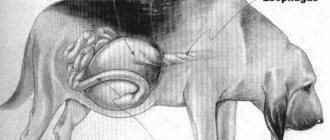
- Anatomy of the spinal column
- Symptoms
- Forms of the disease
- Intervertebral disc disease type I
- Intervertebral disc disease type II
- Intervertebral disc disease type III
- Diagnostics
- Treatment
- Prevention
Discopathy in dogs is an outdated and generalized name for a group of diseases that affect the intervertebral discs in dogs, causing their destruction and, as a result, damage to the spinal cord.
In everyday life and in the literature, the name “disc herniation” is also often found; in the modern classification of neurological diseases, this term is also not recommended for use, because it incorrectly describes the essence and pathophysiology of the process.
Most leading practicing veterinary neurologists consider it most correct to use the term, which is a tracing-paper from the English language - intervertebral disc disease - IVDD.
Understanding the essence of the disease is impossible without understanding the anatomy of the dog’s spinal column and spinal cord.
Anatomy of the spinal column
The spinal column consists of individual vertebrae, which have a complex shape and consist of a body, an arch and processes.
There are five sections in the spine of dogs: cervical ( C
- vertebrae cervicales), thoracic (
T
- vertebrae thoracicae), lumbar (
L
- vertebrae lumbales), sacral (
S
-vertebrae sacrales) and caudal (
Cd
- vertebra caudales).
A dog, like any mammal, always has 7 cervical vertebrae. There are usually 13 thoracic vertebrae, but there may be one more or less (12 or 14). There are usually 7 lumbar vertebrae, but there may be 6 or 8; sacral vertebrae 3 or 4, caudal vertebrae usually from 15 to 25.
Intervertebral discs are cartilaginous pads located between the vertebral bodies throughout the entire spinal column, except from the atlanto-axial joint (between the first and second cervical vertebrae) and the sacral region. There are also intervertebral discs between the caudal vertebrae, but discopathy in this section does not lead to problems with the animal’s health, so it is not an independent medical problem. The main function of intervertebral discs is to act as an elastic shock-absorbing element, absorbing shocks and loads.
The thickness of the intervertebral discs varies from 1.5 to 4 mm. The discs in the thoracic spine are thinner than those in the cervical and lumbar spine.
Anatomically, the disc consists of a gel-like central (pulpous) nucleus and a fibrous (connective tissue) ring. The disc is very strong, as strong as bone.
The nucleus pulposus is a vestige of the notochord. In the intervertebral disc, it is not located in the center, but is slightly shifted upward, towards the spinal canal in which the spinal cord is located.
The spinal cord is located inside a bony canal formed by the processes of the vertebrae and is covered with protective meningeal membranes. Motor and sensory nerve fibers emerge as part of the large spinal nerves in the space between two adjacent vertebrae on each side.
Any compression of the meningeal membranes or roots of the spinal nerves causes severe pain, and compression of the spinal cord itself causes damage to first the superficial (sensitive) and then deeper (motor) nerve fibers, which determines the symptoms of BMPD.
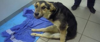
Symptoms of discopathy in dogs
The symptoms that occur with BMPD are associated with compression (squeezing) of the spinal cord by the altered intervertebral disc, which moves upward towards the spinal canal.
The symptoms and degree of neurological deficit directly depend on how severely and “deeply” the spinal cord is affected.
1. The first degree of neurological deficit is manifested in the fact that the dog experiences pain, the gait becomes stiff, the dog refuses typical motor reactions - jumping, running, and does not want to climb stairs. There may be a slight proprioceptive deficit, i.e. decrease in fine skin sensitivity (as a rule, this is difficult to notice with the naked eye and is determined using special tests at an appointment with a veterinarian).
2. The second degree of neurological deficit is manifested by a more pronounced decrease in proprioception (it is noticeable that the dog slightly drags its pelvic limbs) despite the fact that independent movement is possible. There may be no signs of pain.
3. Third degree of neurological deficit: the pelvic (or both pelvic and thoracic, if compression occurred in the cervical region) limbs are paralyzed, but the dog consciously controls urination. Deep pain sensitivity is preserved - the dog responds to a strong painful stimulus (strong compression of a finger of a paralyzed limb with a clamp) by trying to turn around, bite, licking its lips or any other “head” reaction. (NOT withdrawal of the limb - this is a flexion reflex that is not relevant to the assessment of deep pain sensitivity).
4. Fourth degree of neurological deficit: paresis (paralysis) with preservation of deep pain sensitivity, but without urinary control. The bladder can empty itself after fullness, but the dog cannot consciously control the bladder sphincter.
5. Fifth degree of neurological deficit: paralysis accompanied by a lack of deep pain sensitivity (first 24 hours).
6. Sixth degree of neurological deficit: paralysis, accompanied by a lack of deep pain sensitivity, if more than 24 hours have passed since spinalization (the onset of paralysis).
Intervertebral disc disease type I
This type of disease is characterized by rupture of the fibrous ring of the intervertebral disc and the release of altered contents of the nucleus pulposus into the spinal canal. This type of development of the disease is characteristic of dogs of chondrodystrophic breeds, which have a predisposition to early “aging”, dehydration and calcification of the nucleus pulposus of the intervertebral disc.
Chondrodystrophic breeds include:
- dachshunds of all varieties
- beagles
- bulldogs (French and English)
- pugs
- welsh corgi
- basset hounds
- Pekingese
- spaniels
- poodles
- Lhasa Apso
- shih tzu
The disease manifests itself in young and middle-aged animals, usually acutely - symptoms develop within 1-5 days.
The spinal cord can be affected both directly at the level of the spinal disc in which the nucleus pulposus ruptured, and above the bodies of adjacent vertebrae, because The nucleus pulposus “leaks out” and is randomly distributed throughout the spinal canal.
Intervertebral disc disease type II
Type 2 intervertebral disc disease is characterized by protrusion of the fibrous ring into the spinal canal (“bulging disc”), sometimes with extrusion of parts of the nucleus pulposus through the cracks of the ring. Its development is caused by age-related degeneration of the intervertebral disc. Basically, this type is characteristic of non-chondrodystrophic breeds. Manifests chronically with a gradual increase in neurological symptoms from grade 1-2.
Although BMPD type 2 can occur in dogs of any breed, the most predisposed are:
- german shepherds
- labradors
- dobermans
- Rottweilers.
The disease usually manifests itself in older animals. The spinal cord is compressed directly at the level above the intervertebral disc. Very often, protrusions (disc depressions) are multiple.
Types of disease
The types of discopathy are identified based on the specific location of the problem: the cervical or thoracolumbar region. In both cases, there are many similar symptoms of the disease, but at the same time, some differences are noted. In order to correctly diagnose and prescribe adequate treatment, both the doctor and the animal owner must be aware of them.
Cervical
In the cervical spine, the disease can develop very quickly, often affecting young animals up to three years of age. This type is characterized by degeneration of the fibrous membrane of the disc with an increasing process of mineralization of its core, which becomes the cause of extrusion (protrusion of the fibrous ring with a violation of its integrity). The pressure on the spinal cord only increases, so it is not surprising that very soon swelling appears and inflammation of the nerve is noted. The dog tries to turn its head less and seems constrained.
Lumbar-thoracic region
In comparison with the previous option, all destructive processes in the lumbar-thoracic region occur much more slowly, more often affecting older dogs over the age of seven years. Degenerative processes in the spinal cord usually lead to disc protrusion (protrusion while maintaining the integrity of the fibrous ring), and compression of the nerves.
Did you know? The officially registered record for life expectancy in dogs belonged to a terrier mix named Max. He died in 2013 in the United States at the age of 29 years and 5 months.
Often, not one, but two vertebrae are affected, causing severe pain in the animal. One of the most dangerous options is considered to be a lesion of the L5-S1 disc, in which the sciatic nerve is compressed, and the pet cannot sit down, arching its back unnaturally even when moving around the house. Attempts to change body position cause the dog even more pain, causing him to yelp.
Intervertebral disc disease type III
This type of disease is quite rare in both chondrodystrophic and non-chondrodystrophic breeds of dogs, mainly in young animals.
As a rule, symptoms arise abruptly after a significant load, the fibrous ring ruptures, and the nucleus pulposus or part of it is “shot” into the spinal canal at high speed, which leads to severe compression, swelling or rupture of the spinal cord, causing paralysis to occur acutely ( neurological deficit grade 4-5). This type has a poor prognosis.
Causes
Chondrodystrophoid dog breeds - dachshunds, pugs, bulldogs, Pekingese - are prone to discopathy. Dachshunds are at greater risk for this disease. In more than 60% of cases, discopathy is observed in dogs of this breed.
Dogs aged 4 to 7 years are most often affected. Predisposing factors of the pathology are obesity and other metabolic disorders. Less commonly, pathology occurs after spinal injury.
The main cause-and-effect factor in the development of discopathy is cartilaginous metaplasia. The process refers to forms of pathological regeneration, when tissue of one type is replaced by another. This is how fibrous tissue is replaced with cartilage or bone.
Metaplasia develops against the background of chronic inflammation, tissue nutrition or metabolism.
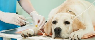
Diagnostics
A preliminary diagnosis is made based on a neurological examination and medical history. A veterinary neurologist determines the degree of neurological deficit and the level of damage - i.e. answers the question of conduction at the level of which segments of the spinal cord is impaired and to what extent.
Further diagnosis consists of neuroimaging methods: this is MRI of the corresponding part of the spinal cord or, if this study is not available, myelography.
Myelography is the injection of a radiopaque substance under the dura mater of the spinal cord. After this, the contours of the spinal cord become visible on a regular x-ray (without contrast, both the spinal cord and intervertebral discs are transparent to x-rays), due to which you can see areas of deformation, compression or destruction (myelomalacia) of the spinal cord.
Thanks to these studies, you can find out exactly which disc is damaged and determine the location of surgical intervention if necessary.
Diagnosis
To confirm discopathy in a dog and prescribe treatment, the doctor must conduct a full examination.
He needs data about when the first signs of the disease appeared and what they were expressed in. Information will be required about the animal’s age, previous illnesses, and whether it had any injuries.
Blood is taken for general and biochemical analysis.
The doctor tests the animal for neurological disorders. Changes in behavior, motor activity and a decrease in the pain threshold indicate spinal cord damage.
Reveal:
- ability to move at a calm pace. At the same time, muscle tone is assessed, whether there is lameness, head position, posture and gait are checked;
- the ability to run, jump, go up and down stairs;
- degree of reflex activation. Deviation from the norm appears as a result of damage to the spinal cord;
- Is there pain sensitivity? Using special clamps, pain is caused on the paws, between the phalanges of the fingers. If the dog only withdraws its paw, the pain threshold is lowered. The normal reaction is to whine and try to bite for protection.
An X-ray examination is performed. It shows the extent of disc damage and how much the spinal cord is involved. In some cases, the study refutes discopathy and points to vertebral neoplasms, destruction of the vertebrae, and mechanical damage to the spine.
A more accurate diagnosis and determination of the specific location of the damage can be made by performing myelography. For this examination, a contrast agent is injected under the membrane of the spinal cord. On an x-ray, it highlights those areas of the spine that are not reflected in a regular x-ray.
The most accurate examination method is computed tomography or magnetic resonance imaging. This examination is expensive and is not carried out in all veterinary clinics.
Treatment
Treatment depends on the degree of neurological deficit.
If we are talking about the initial stages, when there is only pain and impaired movement, conservative (non-surgical) methods are used - painkillers and anti-inflammatory drugs are prescribed and strict rest (limitation of movements) with the help of cellular maintenance for a period of at least 3 weeks. Exact localization of the lesion (data from MRI, CT or myelography) is not required to prescribe conservative therapy.
Often such therapy is enough to prevent further development of the disease and relieve the dog of pain for a period of several months to several years, and with some luck - forever. However, you need to understand that the disappearance of symptoms does not mean that the dog has recovered - if the intervertebral discs are affected by degenerative changes, their “recovery” will no longer occur. If, after a period of cellular dormancy, the dog experiences a relapse, diagnostics are performed to localize the lesion (MRI) and surgical treatment.
If the degree of deficiency is higher than the second (the animal is paralyzed), then, as a rule, symptomatic treatment is not enough; it is necessary to eliminate the cause of compression (compression) of the spinal cord as soon as possible in order to prevent damage to the deep structures of the spinal cord and irreversible changes in it.
The operation performed to provide decompression is called laminectomy and consists of partial removal of the vertebral arch above the site of displacement of the intervertebral disc and extraction of the disc substance from the spinal canal.
Removing the vertebral arch ensures that spinal cord compression does not develop again in the same location, but does not preclude re-displacement of the disc in another (adjacent) intervertebral space - this is uncommon, but occurs in dogs with multiple degenerative changes in the discs.
The earlier the operation is performed, the better the prognosis for surgical treatment. There are isolated cases of recovery of animals operated on with degree 6 neurological deficit, but in general the prognosis at this stage is poor.
Physiotherapeutic rehabilitation methods for spinal patients play an important role in the recovery of animals after surgery.
In addition, if laminectomy is unsuccessful or performed too late, with the help of intensive physiotherapy it is possible to develop the so-called spinal (reflex) walking in a hopelessly paralyzed dog.
How is the diagnosis carried out?
At first glance at a hunched over and tense animal, it is impossible to say unequivocally that the cause of this condition is discopathy, so veterinarians conduct a comprehensive examination, including laboratory tests (primarily biochemical and clinical blood and urine tests), as well as an X-ray examination of the spine.
We advise you to find out what to do if your dog’s ear turns red inside.
To obtain a more complete picture of the condition of the disc, CT (computed tomography) or MRI of the spine can be used, and in their absence, myelography will help determine the location of the narrowing of the spinal canal. This method is considered more dangerous and not as informative as the previous ones, but in small cities there may simply not be another solution. MRI results allow us to determine the location of a specific problem, the volume of prolapsed disc contents, the level of compression and secondary damage to the spinal cord, which is extremely important before performing surgery and drawing up a further treatment plan.
After taking an x-ray, the doctor will be able to assess the degree of reduction in the intervertebral space (signaling the release of the nucleus pulposus), the presence of calcifications in the intervertebral and spinal cavity, the amount of free space between the vertebral processes, which becomes less and less as the disease progresses. When assessing the history, important data will be the presence of a dog’s genetic predisposition to the development of discopathy, the presence of serious injuries in the past and the current stage of development of the disease.
Prevention
Unfortunately, it is impossible to prevent the development of intervertebral disc diseases with absolute reliability.
In a global sense, the main method of preventing degenerative diseases of the intervertebral discs is breeding hygiene - the exclusion from breeding of dogs that have had this pathology and their immediate relatives.
For a particular dog, especially if we are talking about a chondrodystrophic breed, the main and accessible prevention of discopathy in dogs is maintaining good physical shape (developed muscle corset, lack of excess weight).
Author: Kurganskaya (Shuraleva) Natalia Ivanovna
(c) Veterinary center for the treatment and rehabilitation of animals “Zoostatus”. Varshavskoe highway 125 building 1. tel. 8
Signs and symptoms
The severity of the manifestations of discopathy may vary depending on the localization of the pathological process, its stage, and the degree of compression of the nerve roots. At the beginning of the development of the disease, pathological changes may be practically invisible outwardly.
As degenerative changes progress, the dog may develop symptoms of discopathy:
- stiffness of head movements when the cervical spine is affected, the dog tries to move it less,
- the spine is unnaturally curved,
- walking on bent legs, lameness,
- tense, stiff gait,
- due to back pain, the animal does not allow itself to be petted,
- inappropriate reaction to touch,
- abdominal muscle tension,
- uncoordinated movements
- trembling in the body and limbs, twitching of the tail.
Symptoms often increase during periods of active walking and intense exercise. In severe cases of discopathy, the animal may develop paralysis of the limbs, impaired defecation, and involuntary urination.