Gastrointestinal bleeding in a dog
Gastrointestinal bleeding is a fairly common problem in dogs.
It can be a symptom of disease, infection or injury. At the same time, it is not always possible to determine its presence or severity by external signs, since in addition to the pronounced form, there is also a hidden form. In most cases, mild and moderate types are easily treated or go away on their own, but the process still cannot be left to chance.
Moderate blood loss can easily become severe and lead to death or serious complications.
Bleeding in children
Bleeding in children is a very serious reason for going to a medical facility. It will not go away on its own, even if the child vomits blood, and after that behaves normally, plays and asks for food. Before applying, remember if he could have eaten chocolate, hematogen or foods that are colored red (beets, cakes with red dye). Also rule out injuries in the mouth and nose (they are visible to the naked eye).
There are quite a few causes of gastrointestinal tract problems in children. When looking for a diagnosis, doctors first of all pay attention to the age of the child: there are diseases that are most typical for a particular age period:
| Age | Diseases |
| 2-5 days of life | Hemorrhagic disease of newborns - vitamin K deficiency. Characterized by dark, profuse stools 3-4 times a day |
| Up to 28 days of life | Ulcers of the stomach (more often), duodenal ulcers (less often), ulcerative necrotizing colitis of newborns |
| From 14 days to 1 year of life | Duodenal ulcers (more often), stomach ulcers (less often) |
| 1.5-4 months | Intussusception |
| 1-3 years | Juvenile intestinal polyps, Meckel's diverticulum, Dieulafoy's disease, familial colon polyposis (in 5% of untreated children it transforms into cancer by age 5) |
| Over 3 years old | Esophageal varices |
| 5-10 years | Portal hypertension syndrome, ulcerative colitis |
| 10-15 years | Peutz-Jeghers syndrome, when many small polyps are found in the intestines. In this case, the skin, lips, eyelids have a characteristic feature - multiple brown spots |
At any age of a child, starting from the neonatal period, the following may occur:
- gastritis: the cause may be a serious illness, hypoxia (for example, in newborns);
- esophagitis. Most often it occurs in children with shortened esophagus, achalasia cardia, hiatal hernia;
- doubling of the stomach;
- duplication of the small intestine;
- Mallory-Weiss syndrome;
- hiatal hernia;
- eosinophilic gastroenteropathy;
- vascular malformations of the gastrointestinal tract: hemangiomas and vascular malformations.
Diagnostics and emergency care for children is provided on the same principle as for adults.
Causes
The onset of gastrointestinal hemorrhage can be triggered by a gastrointestinal infarction or any disease. It comes from the stomach, esophagus, and various parts of the intestines. The severity depends on how badly the mucous membrane is damaged and which vessels are affected. Ulcers on the arteries are the most dangerous.
The causes of occurrence are:
- Systemic diseases affecting metabolism. Liver disease, pancreatitis, uremia or hyperadrenocorticism.
- Neurology: head and mucous membrane injuries, foreign bodies, intervertebral disc pathologies.
- Use of potent NSAIDs and glucocorticoids.
- Ischemic problems. Volvulus of the stomach or intestines, intussusception, thrombosis of mesenteric vessels.
- Distribution of Pythim and Histoplasm fungi
- Bacterial infections.
- Parasite attacks.
- Parvovirus and coronavirus.
- Tumors of the digestive system.
- Various algal infections.
- External influences. Surgical operations, sepsis, hypovolemia.
- Inflammatory bowel diseases.
- Growing polyps.
- Hemorrhagic gastroenteritis.
Due to the huge range of possible variations in the occurrence of the problem, it is impossible to carry out treatment at home with folk remedies, since even if you manage to get rid of the symptoms for a while, you still cannot eliminate the root cause of their occurrence.
Symptoms
Regardless of where the leak begins, most symptoms will be the same. However, do not forget that hemorrhages can also be hidden. And then the problem can be guessed by indirect signs.
Manifestations of gastrointestinal bleeding include:
- Refusal of food and water.
- Vomiting with blood particles or brown masses.
- Abdominal enlargement.
- Weakness.
- Accumulation of fluid in the abdominal cavity.
- Diarrhea that is brown, black, or scarlet in color.
- Pallor of the mucous membranes.
- Hemorrhages from other openings are possible. For example, through the nose or when urinating.
If such symptoms are noticed, it is necessary to stop feeding and stop giving water, stop taking medications in tablets and urgently seek help from a veterinary clinic, where the primary problem will be identified. You should use therapeutic methods yourself with great caution if you do not know exactly what may have caused the bleeding.
Which medicine is effective?
The causes of thin blood in women and men can be very diverse - from genetic predisposition to a serious illness that provoked a decrease in protein in the blood. The doctor prescribes specific medications depending on the individual characteristics of each patient. It is important to take into account the general state of health, the presence of other pathologies and the person’s age.
Diagnostics
Gastrointestinal hemorrhage can be diagnosed only after determining the root cause and the exact location of vascular damage. Since animals often arrive at the clinic already in serious condition, the initial examination includes a history, physical examination and emergency ultrasound, which will help identify the source of the problem. After resuscitation, all other studies are carried out.
If the patient’s condition is not so serious, then in addition to ultrasound, blood and discharge tests are performed, which show the general condition of the body.
The standard set of studies consists of:
- Tests that determine hidden causes . If the very fact of bleeding is confirmed, then the question arises about the onset of its occurrence, and here doctors will be helped by a whole list of studies of various types: endoscopy, X-ray, ultrasound, complete, general and biochemical blood tests, ACTH stimulation test, etc.
- Tests to confirm the diagnosis for small dogs . If owners of a representative of the dwarf breed come to the clinic with a complaint of progressive anemia, then a full biochemical analysis is carried out to find out the specific cause of what is happening. Very often, such animals experience hemorrhage from the gastrointestinal tract with periodic stops, but then resume again. For a more detailed study, they also take a stool test for hidden traces of blood. Before carrying out it, it is recommended to exclude meat from the diet for 3 days to avoid confusion.
Treatment
The most important thing when treating patients with this diagnosis is to urgently stop the state of shock if the animal has already entered it. Only after this can you begin therapy aimed at stopping the bleeding and healing damaged vessels. The treatment regimen consists of several areas. Necessary:
- Stop the bleeding.
- Replenish its deficiency in the body.
- Relieve pain.
- Improve clotting rate.
- Heal ulcers that have damaged blood vessels.
- Treat the root cause.
The methods used to achieve these goals may vary depending on the individual patient's condition. Transfusion is allowed to restore normal oxygen circulation in the body. Also, in certain cases, it is allowed to use antibiotics if the cause of the disease is infection or bacteria.
If you suspect a stomach ulcer, it is strictly forbidden to give food and water orally, and you should not take pills. All medications and necessary substances and microelements will be delivered to the organs thanks to droppers.
An important role in a successful recovery with a minimum of complications is played by the conditions in which the dog is kept during the recovery period. The dog must be in a warm, clean room, not be subjected to nervous or physical stress, and follow the rules of a special diet.
Forecast
The prognosis for gastrointestinal bleeding is cautious. They depend on the root cause of the disease, the duration and strength of blood flow, the age and physical condition of the patient. Most often, the animal survives provided timely medical assistance is provided.
Mortality rates range from 30 to 45%. In most cases, death can be avoided, but complications from the illness will remain for life. Animals that have experienced this must follow a certain diet throughout their lives and avoid strenuous physical activity.
Source
Urgent Care
Most often, when gastric bleeding develops in newborns, they are still in the maternity hospital (on days 2-5). This increases the chance of providing quality emergency care to such a child.
The basis of treatment is the implementation of hemostasis and replenishment of lost fluid. Algorithm for emergency care for gastrointestinal bleeding in newborns according to clinical guidelines:
- In case of vitamin K deficiency, children are administered intramuscularly at a dose of 100 mcg/kg. As a rule, after 4-6 hours, coagulation parameters return to normal.
- If there is a significant deficiency of the prothrombin complex, they resort to transfusion of its concentrate preparations - Proplex, Octaplex, Konin.
- If hypocoagulation is proven on a coagulogram or the upper limit of normal prothrombin time and aPTT, fresh frozen plasma is prescribed at a dose of 10-15 ml/kg per 30 minutes. It is necessary to repeat the coagulogram to correct the values by additional administration of FFP.
- If there is no effect from the use of FFP, cryoprecipitate is prescribed at a dose of 5 ml/kg over 30 minutes. If no effect is observed, then re-introduce cryoprecipitate at the same dose.
- Platelet mass according to indications is less than 30*10 9 /l at a dose of 1 unit.
- Deficient coagulation factors are prescribed if their deficiency is proven. For example, Ⅷ factor for hemophilia, Ⅸ factor for Christmas disease.
- Red blood cells are administered when there is significant blood loss and a decrease in hemoglobin.
- Children who are not helped by the above remedies are prescribed an exchange blood transfusion, which makes it possible to replenish the missing components of the coagulation system and get rid of decay products.
Key points
- Most of the causes of gastrointestinal bleeding are amenable to therapeutic correction, although in some cases surgical treatment is indicated.
- Gastrointestinal bleeding is an important cause of blood loss anemia in dogs and cats.
- The most common cause of gastrointestinal bleeding described in dogs and cats is ulceration (ulceration) of the gastrointestinal tract.
- the presence of hematomesis and melena allows one to suspect gastrointestinal bleeding, but these signs are not always observed.
- It is reasonable to use gastroprotectors until the cause of gastrointestinal bleeding is confirmed.
- If a patient has gastrointestinal bleeding, the patient’s hemodynamic (cardiovascular) condition should be examined. If there are signs of shock, immediately begin intensive correction of the condition.
- In cats, the typical cause of ulceration is a neoplastic process.
- In dogs, thrombocytopenia should not be ignored as a cause of gastrointestinal bleeding.
- A common cause of ulceration in dogs is the use of NSAIDs and liver disease.
Prevention
In most cases, blood in your dog's stool can be prevented. Key measures that can be taken:
- Anthelmintic treatment. Prevention medications are given once a quarter. If worms are noticed in the feces, deworming is carried out twice with an interval of 10-14 days.
- It is always necessary to monitor the animal's stool - consistency, color, presence of mucus or blood impurities.
- Proper, balanced nutrition with enough fiber. The amount of food should correspond to the weight of the dog. Avoid overfeeding. It is advisable not to give bones often.
- If an animal is predisposed to problems with the digestive tract, it makes sense to immediately transfer the pet to a special diet. They contain easily digestible ingredients and support healthy intestinal microflora.
- While walking, be careful that your dog does not pick up anything from the ground.
- Conduct annual routine vaccination against viral diseases of dogs.
Video:
Introduction
Gastrointestinal bleeding in dogs and cats
is a significant cause of blood loss anemia and is a potentially life-threatening condition for dogs. Rarely described in cats. Bleeding can be acute, chronic, hidden (without visible blood), severe (with a large volume of bleeding, blood). It can be of moderate severity, stop on its own, or a severe, life-threatening condition. Significant bleeding can be detected by physical examination and history. However, you can ignore gastrointestinal bleeding if there are no signs indicating that the source of bleeding - the gastrointestinal tract or a concomitant disease hides this diagnosis. It is important to identify patients with gastrointestinal bleeding in dogs and cats as quickly as possible and initiate treatment to prevent worsening of the condition. This is because even moderate bleeding can progress to a life-threatening condition.
Etiology
Gastrointestinal bleeding
may develop as a result of gastrointestinal infarction or be a complication of a systemic disease, it may originate from the esophagus, stomach, small or large intestine. Many pathological processes can be associated with gastrointestinal bleeding in animals. In general, they can be divided into 3 main categories: diseases causing ulceration, coagulopathies, diseases associated with vascular abnormalities. an animal may have one or more predisposing causes. The most common reported cause of gastrointestinal bleeding in dogs and cats is ulceration of the gastrointestinal tract. The severity of bleeding depends on the degree and intensity of mucosal damage. If erosion damages the underlying artery, the intensity of bleeding depends on the size of the erosion and the diameter of the damaged vessel. Diseases in which ulceration or gastrointestinal tract infection is possible are listed below. The most common causes of ulceration in dogs have been reported to be NSAID use and liver disease.
The neoplastic process is a risk factor for ulcers in cats. These processes described include: mastocytoma, gastrinoma, intestinal lymphosarcoma. An important non-tumor cause that causes gastrointestinal tract is inflammatory bowel disease. Stress ulcers, as a cause of gastrointestinal bleeding, are often described in people who are critically ill. Cases of the development of stress ulcers due to hypovolemia and surgical intervention have also been described in dogs and cats. The validity and statistical significance of stress ulcers in animals has not been confirmed, but their development should be assumed in patients if gastrointestinal bleeding in dogs and cats develops during a hospital stay.
Rodenticide poisoning, disseminated intravascular coagulation syndrome, coagulation factor deficiency (factor XII and prekallekriin factor) and thrombocytopenia are blood clotting disorders associated with gastrointestinal bleeding in dogs and cats. Thrombocytopenia is a common cause of GI bleeding in dogs and should not be ignored. Coagulopathy, as a cause of gastrointestinal bleeding, is less common in cats.
Vascular pathologies due to frequent cases of varicose veins, typical causes of bleeding from the stomach and intestines in humans. In contrast, only a few cases of vascular anomaly have been described in the veterinary literature, and it is an uncommon cause of gastrointestinal bleeding in dogs and cats. This reason should be considered when other, more typical ones have been excluded.
How to check for internal bleeding in a dog
Internal bleeding in animals can be obvious or hidden. In the first case, you can detect it yourself, but only a qualified veterinarian can confirm the diagnosis. If the bleeding is hidden, its independent detection is possible only with careful monitoring of the dog’s condition.
With obvious bleeding, all the fluid enters the gastric cavity or interstitial space. If the problem is related to vascular disorders in the stomach, intestines and lungs, blood appears in the animal’s saliva when coughing. The mass released during vomiting is similar to coffee grounds. In addition, blood is noticeable in stool and urine.
It is much more difficult to detect hidden bleeding. It is necessary to carefully monitor the dog's condition. You should be wary if you notice shortness of breath, bloating, excessive fatigue, decreased activity and pallor of the mucous membranes. Elastic hematomas appear on the bends of the knees. Eyes turn red.
Internal bleeding can be accurately determined by a thorough external examination by a veterinarian, a puncture, and an abdominal x-ray. The latter type of laboratory test detects the presence of fluid in the stomach, but is not able to accurately determine its type.
The puncture allows you to get a more detailed picture of what is happening. Fluid is taken from the animal's stomach for subsequent analysis to determine what type of bleeding is (acute or chronic) and the amount of fluid lost.
Diagnostics may also include:
- Platelet count test.
- Complete blood test.
- Urine and stool analysis.
- Serological analysis.
- Coagulability assay.
- Ultrasound of the abdominal cavity.
- Bone marrow examination.
If necessary, several types of diagnostics can be performed at once. It all depends on the symptoms present, the complexity of the case and the doctor’s recommendations.
History and physical examination
Extensive bleeding, vomiting, diarrhea, or ulcer perforation may result in patients with gastrointestinal hemorrhage presenting in shock due to blood loss, hypovolemia, endotoxemia, or sepsis. When examining such patients, the following signs of shock are revealed: tachycardia, decreased filling or thread-like pulse, cold extremities, lengthening of the SNK, pallor of the mucous membranes. Aggressive therapy is required to relieve shock; searching for the source of bleeding and specialized treatment must be postponed until the hemodynamic state has stabilized.
After initiating antishock therapy, a thorough physical examination and a detailed medical history should be obtained. Hematomesis (vomit that is coffee-colored or bloody), hematochezia (passage of bright red or pure blood with or without stool), or melena (black stool) suggests that the bleeding is coming from the GI tract. However, these signs may not become clinically apparent until the bleeding becomes more severe. Blood in the vomit during bleeding localized in the duodenum may not be visualized if an insufficient (small) volume of the contents of this intestine is thrown into the stomach. But, if bleeding is present, then hematomesis implies ongoing bleeding. Hematomesis and melena can accompany diseases of the nasal cavity and oropharynx, as a result of epistaxis (nosebleeds) and hemoptisis (hemoptysis) and ingestion of blood. These causes of melena and hematomesis should be considered. And finally, black stools can be caused by a diet high in iron, taking activated charcoal, or bismuth. This type of stool should not be confused with melena.
The use of aspirin or other NSAIDs is not uncommon during the history taking. Cases of ulceration and perforation of the gastrointestinal tract have been described in patients of veterinary clinics who received selective cyclooxygenase inhibitors in therapeutic doses. Decreased or lack of appetite in patients receiving NSAIDs should suggest possible complications from the gastrointestinal tract. The use of drugs should be discontinued and the patient examined. If the cause of gastrointestinal bleeding in cats and dogs is thrombocytopenia or coagulopathy, there may be a history of bleeding from other sites, including the nasal cavity or urinary tract. In patients with severe thrombocytopenia, petechiae can be detected on the mucous membranes when examined. Examination of subcutaneous nodules or tumors will identify underlying mastocytomas.
The abdomen (abdomen) should be carefully examined because bleeding from the gastrointestinal tract
may occur and develop asymptomatically, especially chronic bleeding. Palpation of the abdominal wall will allow you to localize the area of pain (weakness, a voluntary or involuntary attempt to defend yourself, or induce vomiting, identify tumors or foreign bodies, determine the enlargement of the abdomen (stretching of the abdominal wall) or signs of the presence of fluid in the abdominal cavity (waves during percussion). In patients with mastocytoma or other neoplasias, liver disease may show hepato or splenomegaly. At the initial examination or during resuscitation, a thorough rectal examination should be performed to identify melena or pure blood, tumors or foreign bodies. Although bleeding from any part of the gastrointestinal tract can be serious, bleeding from the upper tract tends to be more severe. In addition, the etiology, diagnostic techniques, and treatment of upper and lower GI bleeding are different. Thus, it is important to determine the area of bleeding. The presence of hematomesis or melena suggests upper GI bleeding. However, it should be remembered that blood remains in the gastrointestinal tract for some time and it is not necessarily this location that determines the color. Increased transit time and retention in the large intestine lead to the appearance of melena, which is associated with damage to the lower gastrointestinal tract. Hematochezia usually reflects bleeding from the colon, rectum, or anus. However, acute severe intestinal bleeding acts as a laxative, which significantly reduces the transit time. This may result in pure blood in the stool due to significant blood loss from the upper GI tract.
Diagnostics
Bleeding in cats and dogs has been proven
when the source of bleeding is identified. In patients with signs of shock, during correction of the condition and search for hidden causes, a minimum number of emergency blood tests are performed, namely hematocrit, solids, blood nitrogen (BUN), glucose, if possible, pH, lactate and electrolytes. If hemoabdomen or septic peritonitis is suspected, emergency ultrasound, laparocentesis and diagnostic peritoneal lavage are indicated, which can be performed during the initial correction of the patient's condition. Once resuscitation has been initiated or the patient's condition has been stabilized, other diagnostic modalities should be considered.
Tests to Confirm Gastrointestinal Bleeding in Small Pets
If physical examination and history do not reveal obvious symptoms of gastrointestinal bleeding in dogs and cats, hematological and biochemical studies are performed, the results of which may suggest bleeding. Anemia of unknown etiology may lead to the assumption of bleeding from the gastrointestinal tract; it is described that microcytic hypochromic anemia (iron deficiency anemia) develops as a result of chronic blood loss in the gastrointestinal tract. Because iron deficiency anemia takes time to develop, a more typical consequence of recent bleeding is normocytic normochromic anemia. A high urea-creatinine ratio (greater than 20) has been described in gastric and intestinal bleeding, especially if the bleeding occurs from the upper gastrointestinal tract. This is due to bowel movements and intestinal absorption of proteins (may increase the level of digested and absorbed protein), including digested blood, into the circulatory system (blood). Massive intestinal bleeding, as described, has little effect on urea levels. However, once protein metabolism is activated as a result of illness (fever, infection, fasting or use of glucocorticosteroids), the urea-creatinine ratio may also increase, which must first be concluded to be the cause of gastrointestinal bleeding in dogs and cats.
In doubtful cases, a fecal occult blood test is performed. Most tests are based on the peroxidase activity of hemoglobin. Although the test is useful in detecting occult bleeding, it is possible to obtain a false positive result. This is influenced by the following factors: diets that include red meat or foods with high peroxidase activity, such as fruits, fish, vegetables. The result is also affected by bacteria with peroxidase activity present in the gastrointestinal tract. This must be kept in mind when receiving test results based on chemical characteristics.
It should be recommended to exclude meat from the animal’s diet for at least 72 hours. On the other hand, a negative occult blood test excludes significant GI bleeding. When significant gastric bleeding is suspected but not confirmed, a nasogastric tube is inserted and the gastric contents are aspirated. This will help confirm and determine the location of the bleeding. Although the procedure can cause discomfort and falsely described - negative results.
Causes of gastrointestinal condition
There are so many causes of acute gastrointestinal bleeding that they are divided into two classifications. One of the classifications designates the type of causes, the second - the causes depending on the location in the gastrointestinal “tube”.
So, depending on the type of reasons, gastrointestinal tract can be caused by:
- Inflammatory, erosive and ulcerative formations of the gastrointestinal tract, as a result of which the vessels feeding one or another structure are “corroded”. Not all of these pathologies occur due to poor diet or Helicobacter pylori infection. Erosive-ulcerative lesions occur with any serious illness (this is called stress ulcers). They are caused by burns from strong alcoholic drinks, acids and alkalis, drunk by mistake or intentionally. Erosion and ulcers also often occur as a result of taking painkillers and glucocorticoid hormones.
- Tumors of the gastrointestinal tract of any degree of malignancy.
- Wounds and injuries of the gastrointestinal tract.
- Blood clotting diseases.
- Increased pressure in the vessels of the gastrointestinal tract. This mainly happens only with portal hypertension syndrome caused by cirrhosis, blood clots in the portal vein or compression from the outside.
Depending on the location, bleeding from the upper sections (up to the end of the duodenum) and bleeding from the lower sections (starting from the small intestine) of the gastrointestinal tract are distinguished. The upper sections suffer more often: they account for about 90% of gastrointestinal tract infections, while the lower sections, accordingly, account for a little more than 10% of cases.
If we consider the frequency of damage to individual organs, then bleeding from the stomach is every second gastrointestinal tract, bleeding from the duodenum occurs in every third case. The colon and rectum are every 10th bleeding, the esophagus is every twentieth. The small intestine in adults rarely bleeds - in 1% of cases.
The causes of gastrointestinal tract from the upper gastrointestinal tract are:
- erosive esophagitis, whose main cause is ingestion of acids or alkalis;
- erosive and hemorrhagic gastritis, including those caused by taking painkillers;
- peptic ulcer of the gastric or duodenal localization;
- increased pressure in the veins of the esophagus (portal hypertension syndrome). It develops with cirrhosis of the liver, blood clots in the hepatic or other veins communicating with the portal vein, compression of the portal vein at the level of the heart - with constrictive pericarditis or at any other level - with tumors and scars of nearby tissues;
- penetrating wounds of the chest or upper abdomen;
- Mallory-Weiss syndrome;
- stomach polyps;
- injuries to the esophagus or stomach caused by foreign bodies or hard (metal) medical equipment during examination;
- bleeding from diverticula (“pockets”) and tumors of the esophagus, stomach or duodenum;
- hiatal hernia;
- aorto-intestinal fistulas;
- injuries of the biliary tract (mainly during operations and manipulations), in which blood along with bile enters the duodenum.
The causes of gastrointestinal bleeding from the lower sections are:
- blunt abdominal trauma;
- abdominal wounds;
- tumors;
- thrombosis of mesenteric vessels;
- infection with worms;
- increased pressure in the veins of the rectum, which is caused by portal hypertension, which has the same reasons as in the case of the esophagus;
- nonspecific ulcerative colitis;
- Crohn's disease;
- anal fissures;
- haemorrhoids;
- diverticula;
- infectious colitis;
- intestinal tuberculosis.
The causes of gastrointestinal bleeding, which can cause bleeding from any part of the gastrointestinal tract, are vascular damage due to:
- systemic lupus erythematosus;
- vitamin C deficiency;
- periarteritis nodosa;
- atherosclerosis;
- Rendu-Osler disease;
- rheumatism;
- congenital malformations, telangiectasias and other vascular malformations,
- coagulation disorders (eg, hemophilia);
- decreased platelet levels or abnormalities in their structure (thrombocytopathy)
In addition to acute bleeding, there are gastrointestinal tract infections of a chronic nature. This means that in a certain location there are damaged vessels of small caliber, from which small, non-life-threatening volumes of blood periodically leak. The main causes of chronic bleeding are stomach and duodenal ulcers, polyps and tumors.
Tests to identify hidden causes
Once gastrointestinal bleeding in dogs and cats is suspected or confirmed, a search for an underlying cause should be undertaken: coagulation tests, complete blood count, routine chemistry profile, electrolytes, ACTH stimulation test, imaging (x-ray, ultrasound), endoscopy.
A coagulation test will help identify coagulopathies such as rodenticide poisoning or clotting factor deficiency. An increase in blood clotting time, which can be detected by testing, is not in itself the cause of gastrointestinal bleeding in cats and dogs, but can significantly aggravate the condition. The platelet count is important because a common cause of severe gastrointestinal bleeding in dogs is immune-mediated thrombocytopenia. An increase in hematocrit in patients with acute hemorrhagic and relatively normal protein concentrations is suggestive of hemorrhagic gastroenteritis.
Taking into account that the confirmed cause of ulceration of the gastrointestinal tract is liver and kidney diseases, special attention should be paid to the biochemical markers of these diseases (alkaline phosphatase, ALT, AST and bilirubin - for liver diseases; urea, creatinine, phosphorus - for kidney diseases). It is necessary to examine the level of electrolytes and perform an ACTH stimulation test, since the confirmed cause of GIB is hypoadrenocorticism. Research is carried out if no other causes of bleeding are found. A stool smear, culture, and test for parvovirus will confirm the suspicion of an infectious disease. Measuring gastrin levels is recommended if ulcers recur and do not respond to therapy.
Radiography will reveal foreign bodies, tumors, or free air in the peritoneal space. The cause of pneumoperitoneum can be assumed to be gastrointestinal perforation if the patient has not undergone abdominal surgery in the recent past. Although contrast radiography will reveal mucosal defects as the cause of gastrointestinal bleeding in cats and dogs, in most cases it can be replaced by ultrasound or endoscopic examination. Ultrasound can identify tumors and foreign objects accompanying gastrointestinal ulcers, if any. The use of ultrasound to recognize (define) ulcers is described. The study allows you to visualize the structure of the intestinal wall, its thickening, defects and craters. If serial testing is carried out (over time), it is possible to confirm changes that occur in response to therapy or the need for surgery in some cases. The use of ultrasound to identify ulcers in cats has also been described.
Endoscopy with greater accuracy (sensitivity) will allow you to determine the location of bleeding or ulcers in the upper gastrointestinal tract. Before the study, the patient's condition must be stabilized. The study often allows you to establish a diagnosis, assess the prognosis, can be therapeutically useful (removal of foreign bodies), and finally, during the study you can directly visualize the mucosa, obtain material for biopsy and culture, which may be necessary to determine damage or infection (for example, tumors, inflammatory bowel disease, protothecosis Disadvantages of endoscopy include the need for anesthesia, limitation to the proximal GI tract and colon, potential increased bleeding, and the possibility of iatrogenic ulceration.
If all of the above diagnostic methods do not allow us to establish the cause of ongoing gastrointestinal bleeding, then diagnostic laparotomy, scintiography with technetium-labeled erythrocytes and arteriography are performed. Using scintiography, you can determine the source of bleeding in dogs, and arteriography will allow you to establish vascular pathology of the gastrointestinal tract.
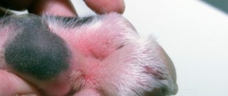
Blood in stool: why it happens
Hematochezia is a pathology characterized by the presence of unchanged blood in the stool. This indicates soft tissue injuries in the lower intestine. If the feces are dark, to an intense black color, we are talking about bleeding in the upper intestines or stomach, the blood in this case is partially digested.
List of reasons for bloody stool in dogs:
- damage to the capillaries in the rectum and colon, blood comes out along with particles of undigested food;
- damage to the anus and intestinal walls with sharp objects (chicken bone fragments);
- inflammation of the gastrointestinal tract, proctitis;
- acute viral infections that damage the mucous membrane (parvovirus, gastroenteritis);
- polyps, other neoplasms on the walls of the intestine, which are often injured or degenerate (malignant forms);
- blood clotting disorder;
- ulcers, erosions along the entire gastrointestinal tract.