The rectum in animals is the final section of the large intestine and is responsible for the evacuation of contents to the outside. Veterinarians rarely diagnose organ prolapse (rectal prolapse) in domestic cats. At the same time, there is a predisposition to the disease in young and aging individuals.
There are many causes of rectal pathology - from helminthic infestations to malignant tumors. The pet owner should know not only the symptoms of the disease, but also methods of helping a sick animal.
Causes of rectal prolapse in cats
In veterinary medicine, the etiological factors of rectal prolapse have been studied quite well. The main causes of rectal prolapse in domestic cats include:
- Chronic constipation. Hard feces lead to overstretching of the wall of the large intestine and weakening of the muscle layer. Constant straining of the animal during defecation leads to prolapse of the anus.
- Diarrhea. The cause of rectal prolapse is often profuse diarrhea. This is due to disruption of the small and large intestines, irritation of the mucous membrane of the digestive tract.
- Feeding your pet dry food. Such compositions, along with a violation of the drinking regime, provoke injury to the mucous membrane of the intestinal tube and, as a result, prolapse of the anus during the act of defecation.
- Chronic diseases of the large intestine - colitis, proctitis - are a common cause of rectal prolapse in animals. Inflammatory processes affect the muscular layers of the anal sphincter, which leads to prolapse of the final section of the large intestine.
- Helminthic infestations. Especially often, helminths cause prolapse of the anus in young animals up to six months of age. The parasites themselves and the toxic products of their vital activity cause irritation of the mucous membrane of the large intestine, which leads to straining and severe tension of the anal sphincter muscles.
- Injuries, perineal hernia, rectal administration of irritating drugs are common causes of prolapse in domestic cats.
- Prostatitis and prostate adenoma in males also lead to rectal prolapse. In females, the cause of the disease is often pathological childbirth, excessive straining during labor.
- Diseases of the urinary system provoke chronic tension in the anus with frequent urination.
- Neoplasms and foreign objects in the preanal area can also lead to rectal prolapse.
- Injuries and tumors of the spinal cord also provoke pathology in furry pets.
Veterinary experts note that most often the pathology is detected in young pets under the age of one year, as well as in aging cats after 10 years. No breed predisposition to the disease was noted.
We recommend reading about uterine prolapse in a cat. You will learn about the causes of uterine prolapse, types of prolapse, symptoms, and treatment from a veterinarian. And here is more information about umbilical hernia in cats.
Diagnostics: basic methods
At the first signs of illness, you should consult a doctor.
If a polyp is discovered or your cat’s rectum has prolapsed, you should see a veterinarian in order to begin treatment for the pathology in a timely manner. If there is a problem, a specialist examines the damaged area and assesses the severity of the disease. It is important to determine whether the disorder can be overcome conservatively or whether surgery will be required. To make a final diagnosis, the cat is prescribed the following diagnostic procedures:
- general and biochemical blood tests;
- laboratory examination of stool;
- colonoscopy;
- X-rays of the abdominal cavity;
- Ultrasound diagnostics of the gastrointestinal tract of a cat.
Signs and symptoms of the problem
Excessive straining for any of the above reasons leads to increased pressure in the animal’s intra-abdominal cavity. The rectum is pushed outward with eversion of the mucous membrane. Often, due to the weakness of the ligamentous apparatus that supports the large intestine, eversion does not occur, and the mucous membrane is screwed into the prolapsed part of the intestine.
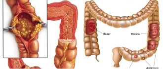
Anatomy of the gastrointestinal tract in a cat
The anal sphincter compresses the prolapsed area. Drying, mechanical damage and microtrauma of the organ are observed. Swelling of the prolapsed rectum develops. This leads to severe pain. In the strangulated part of the organ, blood circulation is disrupted and it becomes infected. If timely assistance is not provided, rectal necrosis and death are possible.
In veterinary medicine, it is customary to distinguish between complete and incomplete prolapse. Complete prolapse is the prolapse of the entire rectum; with incomplete prolapse, only a section protrudes outward. Often, cats also experience internal (hidden) prolapse, which is intussusception without visible prolapse.
Regardless of the type of rectal prolapse, the owner will observe the following clinical signs:
- Problems with going to the litter box. When an animal goes to the toilet, it experiences pain, and therefore the act of defecation is accompanied by moaning, groaning, and meowing.
- Visually, the owner observes the prolapsed rectum in the form of a protrusion from the anus of the animal. Cats do not have hemorrhoids, so any protrusion in the anal area should immediately alert household members.
The appearance and nature of this protrusion depends mainly on the duration of the prolapse and the type of prolapse (complete, incomplete). If the pathology is fresh, then a dark red color of the intestine is observed. The prolapsed part may bleed. If several hours have passed after the prolapse, the prolapsed organ is pinched by the anal sphincter, and a bluish color is observed.
Black color indicates that the necrotic process has begun. Often in advanced cases, ulcers are found.
- The sick animal experiences pain, moves little, and licks the anus area.
The bluish color of the prolapsed organ should cause particular concern to the owner. Veterinarians strongly recommend urgently showing a sick animal to a specialist, and under no circumstances self-medicate.
If rectal prolapse is detected in a cat, the owner should wash the prolapsed intestine with an antiseptic solution and apply a sterile bandage. The animal should be placed on clean bedding and transported in a carrier to a medical facility.
What does it look like?
The two most important symptoms:
- painful attempts to empty the intestines, accompanied by restlessness of the pet, meowing, and subsequent attempts to lick under the tail;
- a pink-crimson “bump” 1 cm or more long appears under the tail, which does not disappear anywhere and does not retract.
Cats don't have hemorrhoids! Any protrusion in the anal area should attract the attention of the owner and serve as a reason to take the cat to the veterinary clinic!
If the functions of the sphincter are not impaired, then the prolapsed section of the intestine can be pinched, the blood circulation is blocked, and the risks of its death (ulcers, necrosis) increase.
Degrees of loss:
- I – only with strong attempts, but independent retraction is observed;
- II – after bowel movement, the prolapse does not reduce, but the protrusion is not strong and it is enough to simply push it back with your hands;
- III – prolapse is observed with any strain on the cat, be it the intestines or, for example, the bladder;
- IV – the rectum falls out even at rest, when walking.
Diagnosis of the condition
The diagnosis of rectal prolapse is made by a veterinarian based on a clinical examination. During the examination, the doctor determines the age of the pathology and the type of rectal prolapse. Based on the visual picture, treatment tactics for the furry patient will be determined.
To determine the cause of the pathology, the pet will be prescribed a series of diagnostic measures. In order to exclude helminthic infestation, a scatological examination is carried out. To detect tumors and foreign objects, X-ray and ultrasound examinations and colonoscopy are used.
Treatment of side and associated symptoms
Cat and medicine for worms.
- Mild laxatives are prescribed for constipation . It is acceptable to add one or two drops of vegetable oil to food. Vaseline oil is used, but before use, consult a doctor. The dose is calculated purely individually, and taking it together with vegetable oil is strictly prohibited.
- Infection with helminthic infestations is treated by prescribing broad-spectrum anthelmintics: polyvercan, dirofen, profender, troncil.
- The cause in the form of diseases of the genitourinary system is eliminated using highly targeted drugs.
Treatment of rectal prolapse in a cat
The choice of therapeutic method for rectal prolapse depends on the duration of the pathology, the type of prolapse and the general condition of the animal.
Therapeutic approach
Conservative treatment methods, as a rule, are effective only in the first hours after the development of the pathology and when the provoking factor is eliminated. In this case, the veterinarian, after preliminary anesthesia (most often sacral anesthesia is used, in rare cases general anesthesia), assesses the condition of the rectum and straightens it.
If no signs of necrosis are observed, then the prolapsed intestine is treated with disinfectant solutions.
The organ is often lubricated with novocaine ointment, as well as ointments that reduce swelling and inflammation (Ultraproct, Proctosan), after which the intestine is inserted into the anus.
In order to prevent relapse, the surgeon places a so-called purse-string suture around the anal sphincter. A special method of applying suture material does not interfere with the act of defecation and prevents recurrent rectal prolapse.
The animal is prescribed painkillers, anti-inflammatory and decongestant drugs. The suture is removed after 2 - 4 days. Eliminating the root cause of rectal prolapse plays an important role in curing your pet.
Operation: performance, after care
If there are signs of necrosis of the prolapsed organ, its ruptures, or repositioning is difficult due to late treatment, the veterinarian considers the issue of rectal resection. Removing the damaged part of the organ is the only chance to save the animal’s life when the necrotic process has begun.
The operation is usually performed under general anesthesia.
The veterinarian selects the most effective method of operation in a particular case (Olikov, Frick, Minkin method, resection using a test tube). The essence of any of them is to remove the necrotic part of the intestine and suturing the remaining fragment to the anus.
Surgery for rectal prolapse in cats
In the postoperative period, the animal is prescribed analgesics and a course of antibacterial therapy. Postoperative care involves wearing a protective diaper bandage. To avoid infection, the pet must be kept in a clean, dry and warm room.
Particular attention is paid to the diet of the operated cat. Dry food is completely excluded. Feeding should be frequent, small portions. The formation of constipation and gas in the animal is not allowed.
We recommend reading about intestinal problems in cats. You will learn about the causes of constipation in cats, signs of constipation, treatment and prevention. And here is more information about the causes of feces with blood in a cat.
How does it manifest?
Treatment at home without doctor's advice can lead to worsening of the condition.
It is difficult not to detect symptoms of a prolapsed rectum in a cat, since the pathology greatly affects the behavior and condition of the pet. Veterinarians do not recommend that owners try to straighten a part of the internal organ themselves, since careless movements will lead to serious consequences. When the rectum protrudes from the anus, the kitten and adult pet are worried about the following symptoms:
- difficulties with bowel movements;
- excretion of feces with blood impurities;
- restless behavior of the animal;
- frequent meowing, which intensifies when going to the litter box;
- Licking the area near the anus.
| Stage | Peculiarities |
| I | Prolapse occurs only during bowel movements |
| The rectum, which emerges from the anus, has a cylindrical or spherical shape | |
| Reduction can be done independently at home | |
| II | Most of the organ is sticking out, but it is still possible to straighten it with your hands |
| III | The problem occurs with any increase in intra-abdominal pressure in a cat |
| It is impossible to return the rectum to its place on your own | |
| IV | Even with a loud meow or light jump, part of the internal organ is noticed in the anus |
Preventing rectal prolapse in cats
In order to prevent the occurrence of a dangerous disease in pets, veterinary experts recommend that owners adhere to the following rules:
- Treating a cat against worms
The diet should be balanced and not lead to chronic constipation and diarrhea. If necessary, on the recommendation of a doctor, you can give your cat probiotics that improve digestion processes. - Regularly treat the animal against helminths. It is recommended that you first undergo a stool test to identify the type of parasites and, based on the results of the examination, deworming is carried out on the recommendation of a doctor.
- Compliance with the scheduled vaccination schedule.
- Treatment of concomitant diseases: proctitis, prostatitis, genitourinary diseases.
- Preventing animals from swallowing foreign objects.
Rectal prolapse in domestic cats is a dangerous disease that, if not treated promptly, can cost the pet’s life. If the rectum prolapses, the owner must urgently take the animal to a specialized facility. In the early stages, it is possible to reduce the prolapsed organ; in advanced cases, resection of necrotic areas is necessary. Postoperative care requires a strict diet to prevent constipation.
What is the risk of injury?
Rectal prolapse is not a harmless condition for a pet, especially if we are talking about a kitten, in which all processes in the body occur much faster than in an adult.
- If you notice symptoms of hair loss in your animal, you should immediately contact your veterinarian. The matter does not require delay, because the prolapsed appendage, exposed to air and the external environment, dries out, becomes sensitive, the mucous membrane becomes covered with microcracks, into which bacteria can enter and cause inflammation.
- In addition, the intestinal segment compressed by the sphincter swells, its blood circulation is disrupted, and in advanced cases, tissue necrosis may develop.
- If the prolapsed appendage becomes infected, sepsis can occur, which leads to the death of the animal.
- The pain that occurs when a prolapsed intestine becomes infected moves to the abdominal area and can cause depression in the pet’s respiratory activity.
Important! Since cats have a much lower pain threshold than humans, it is not always possible to determine the degree of pain by their behavior. Therefore, when the first signs of the disease appear, it is important to immediately show the animal to a doctor.
Mortality from painful shock as a result of prolapse of the appendage of the rectum if assistance is not provided to the cat in a timely manner is very high.
Veterinary clinic of Dr. Shubin
Description
Rectal prolapse is a protrusion or inversion of the rectal mucosa through the anus (anus).
Rectal prolapse can be complete or partial. With incomplete prolapse, only the mucous membrane of the rectum falls out, and this does not occur along the entire circumference of the anus. In complete rectal prolapse, all layers of the rectum fall out around the entire circumference of the anus. The degree of rectal prolapse increases with ongoing tenesmus, and can vary from a few millimeters to several centimeters. After the intestines exit through the anus, the tissues become swollen, which prevents their spontaneous return to the pelvic canal. Prolonged exposure leads to injury to the prolapsed section of the rectum, bleeding, drying out and death of sections of the intestine. Synonyms: rectal prolapse, anal prolapse, anal prolapse.
Causes
In general, there are two peaks of rectal prolapse in cats and dogs: the first peak occurs at a young age and the most common cause is internal parasites and various forms of intestinal inflammation; the second peak is observed in middle-aged and elderly animals, more often observed in dogs, and this is associated with the development and surgical correction of a perineal hernia. However, any condition accompanied by an increased urge to defecate (tenesmus) can lead to rectal prolapse. Below are the main pathologies that can lead to the disease: • Intestinal parasites; • Intestinal inflammation; • Intestinal foreign bodies; • Intussusception; • Pathological labor (severe contractions, dystocia); • Urolithiasis and inflammation of the bladder; • Constipation; • Weakness of the anal sphincter; • Prostate diseases; • Perineal hernia and more often as a result of surgery to correct it; • Some birth defects.
Factors that predispose to the development of the disease include weakness of the connective tissue or muscles around the anus, uncoordinated peristalsis (a combination of intestinal contraction and relaxation of the anus), and inflammation with swelling of the rectal mucosa.
Clinical signs
Rectal prolapse occurs in both cats and dogs, without any breed predisposition. However, there is an opinion that the disease occurs more frequently in Manx cats due to breed weakness of the anus muscles. In the vast majority of cases, prolapse occurs in young animals. When collecting a medical history of rectal prolapse, the animal often describes various episodes of frequent and increased bowel movements; this can occur with many of the diseases described above.
When examining an animal by a veterinarian, rectal prolapse is determined by the naked eye; the disease has a characteristic appearance - masses protruding from the anus, ranging in color from pink to black (depending on how long ago the prolapse was). As mentioned above, the degree of rectal prolapse can range from a few millimeters to several centimeters.
Diagnostics
The appearance of masses protruding from the anus may be the same in two diseases - ileocolic intussusception and rectal prolapse. To distinguish between these diseases, the veterinarian inserts a thin, blunt object (probe) between the anus and the prolapsed masses - with intussusception, the probe easily passes into this gap; with prolapse of the rectum, stable resistance is felt.
Due to the wide variety of causes of rectal prolapse, the veterinarian may offer the owner some other diagnostic tools to identify underlying diseases (eg laboratory tests, radiographic examination, ultrasound).
Treatment
Treatment of rectal prolapse depends on factors such as: the degree of prolapse, the duration of the process, recurrence after treatment and the possibility of correcting the underlying diseases. There are the following main methods for correcting rectal prolapse: manual reduction with the application of a purse-string suture; amputation of prolapsed intestine; colopexy (suturing the colon to the abdominal wall). In each case, the veterinarian chooses one or another treatment method after discussion with the animal owner.
Manual (manual) reduction with a purse-string suture
This treatment method is indicated in acute cases of rectal prolapse with minimal swelling and soft tissue damage. The animal, under anesthesia, is treated with various solutions (saline solutions, gel) to reduce the volume and facilitate reduction. After manual reduction, a purse-string suture is placed around the anus, the veterinarian selects its diameter based on two main parameters: the free passage of feces through the opening; preventing recurrent rectal prolapse. The use of an epidural may facilitate reduction and reduce the likelihood of increased bowel tenesmus. After the purse-string suture is placed, the animal is prescribed a low-fiber diet.
Many patients have a good response to the above-described manipulations, and most return to normal if the underlying diseases are corrected (see above).
Amputation of a prolapsed section of intestine
This method of surgical treatment is used more often in chronic cases, when it is either impossible to achieve manual reduction of the prolapsed area, or the prolapsed area becomes non-viable. When amputating a prolapsed rectum, a non-viable section of the intestine is removed from the animal under anesthesia and an anastomosis is performed (connection of living segments). After successful anastomosis, the surgical site is reduced to a physiological location and a purse-string suture is applied to the anus (not always) for a period of 1-2 days to avoid recurrent prolapse. The purse string suture is removed after the required time; the sutures on the intestine itself are not removed. After amputation, the animal may be prescribed certain laxatives.
Colopexy
Colopexy is the suturing of the colon to the wall of the abdominal cavity. The goal of this operation is to create a permanent fusion between the serosa of the colon and the abdominal wall to prevent caudal (backward) displacement of the large intestine. The most common indication for colopexy is recurrent rectal prolapse.
Colopexy can be performed laparoscopically, but our veterinary clinic lacks both the necessary equipment and experience in performing this type of intervention. Our main method of colopexy is open access through the abdominal cavity and the operation is performed under visual control. For colopexy, two slightly different techniques can be used; according to the literature, their effectiveness is approximately the same.
After the above interventions, work should be carried out to identify and correct the likely underlying causes. Also, postoperatively, the use of various painkillers and microenemas with barium or kaopectate can help to prevent tenesmus.
Forecasts
Incomplete prolapse can resolve spontaneously (without any intervention). In case of complete prolapse of the rectum without manual reduction or other types of correction, the prognosis is unfavorable, the exposed intestinal mucosa is injured, and eventually its death and the development of secondary blood infection (sepsis) develop. With various types of treatment for rectal prolapse, the prognosis is closer to favorable.
Photo 1. A young cat was taken by animal rights activists for an appointment at a veterinary clinic; rectal prolapse is clearly visible under the tail; the probe does not go far.
Photo 2. View of the cat after surgery in a veterinary clinic, the large intestine was returned to its original state, the colon was sutured to the abdominal wall from the inside (colopexy) to avoid recurrent prolapse.
Photo 3. A 3-month-old kitten with rectal prolapse at an appointment at the veterinary clinic.
Photo 4. Applying a purse-string suture to the kitten from photo No. 3, the staff of the veterinary clinic corrected the prolapsed section of the intestine, an insulin syringe was placed in the opening of the anus, around which a nylon suture was tightened.
Veterinary clinic of Dr. Shubin, Balakovo
Diagnostics
Rectal prolapse is a serious problem that, without timely and proper treatment, can lead to the death of the animal. Even if, after partial prolapse, the intestine retracts itself and falls into place, the animal still needs to be shown to a doctor.
Until the cause of the prolapse is eliminated, the animal’s condition will begin to deteriorate. Soon the loss will be complete.
Immediately after the incident, the protruding intestine must be treated with an antiseptic solution to avoid infection. As a last resort, the fallen part can be washed with clean boiled water.
Afterwards, you need to apply a sterile bandage, put the cat in a carrier and immediately go to the veterinary clinic. If the intestine turns blue or black, you cannot wait a minute - the animal needs urgent help.
A visual examination is required to make a diagnosis. Based on the appearance of the intestine, the doctor will make a conclusion about how long ago the prolapse was. It is also important to understand whether the vessels are pinched or whether there are injuries or ruptures in the intestine.
Depending on the stage at which the pathology is, the doctor will prescribe appropriate treatment. In addition, the doctor must determine the cause of rectal prolapse. It is not easy. The animal will have to undergo a series of tests.
- First of all, you will need to have your stool tested to rule out infection with worms.
- A complete blood count may also shed light on the cause.
- Ultrasound examination, X-ray and colonoscopy will help to exclude diseases of internal organs and the appearance of tumors.
Correct treatment
With timely treatment, the problem of rectal prolapse can be solved with minimal difficulties.
Conservative
Conservative treatment is appropriate in mild cases of the disease. It consists of manual reduction of the appendage by a veterinarian and fixative therapy aimed at eliminating the causes of the disease (adjusting a diet, treating helminthiasis, antibacterial therapy).
If you notice that your pet has something sticking out of its anus, do not delay visiting the doctor.
Operational
A quick solution to the problem is resorted to in several cases.
- If the loss recurs. In this case, laparoscopic surgery is performed to attach the intestine to the abdominal wall.
- If the situation is advanced and the appendix is not viable (has manifestations of necrosis), then it is removed surgically. The remaining part of the intestine is sewn into the anus.
We invite you to read: When does a cat’s milk come in: normal and pathological conditions
After surgery, the cat is prescribed an antibiotic.
Any of the operations in this case is performed under anesthesia, the type of anesthesia is determined by the veterinarian.
Important! Do not under any circumstances try to straighten the intestinal appendix at home; this may cause injury to the intestine and cause bleeding.
Inflammation of the anus in a cat: causes, symptoms and treatment methods – Pets Pets
What are the general symptoms of this pathology? In general, there is nothing complicated here, and even a person far from veterinary medicine can determine the presence of the disease.
So, the most characteristic signs:
- The animal is very heavy, with visible tension and groaning, walking on the tray “at large.”
- You can see a “bump” or a “tube” clearly protruding from the anus. Note that in mild cases the tumor is not always visible. The rectum protrudes only during bowel movements.
- Excessive licking of the anal area and genital area.
But the main symptom, of course, is the presence of a protruded, swollen rectum. In appearance, it looks like a swollen, stretched bubble. The longer the organ is in an external position, the more hyperemic (reddened) and swollen its shell becomes.
Let us remind you once again that constant bulging of a segment of the rectum is characteristic only of severe, advanced cases of prolapse. If something pinkish protrudes from the anus during a bowel movement, take your pet to the vet immediately. This disease is much easier to cure in the earliest stages.
Like any disease, this disease primarily causes anxiety in the animal. This is especially noticeable before and after defecation. A cat can constantly lick the anus area, rub against various surfaces, and “ride” on its butt.
An alarming symptom may be the appearance of blood from the anus.
The disease can manifest itself in different ways, depending on the stage of development.
- The first degree is manifested by the protrusion of the appendage at the time of defecation, then the appendage is automatically set back.
- The second degree is characterized by the fact that after defecation the intestine does not reduce itself; manual reduction is necessary.
- The third degree is manifested by the fact that prolapse occurs with any attempts by the animal.
- When the disease develops to the fourth degree, the intestine falls out even when walking.
The first symptoms may also indicate other pathologies (hernia, hemorrhoids, anal fissures), so an accurate diagnosis can only be made by a veterinarian after an examination.
We suggest you read: How to get rid of the smell of cat urine in an apartment at home, how to remove the smell of cat urine in an apartment
Diseases of the anal sacs in cats.
Anal sacs are glands located on each side of the anus. They are scent glands and produce small amounts of dark, odorous fluid that is usually expelled during bowel movements. Anal sac diseases begin as a result of blocked or infected glands. Abscesses or cancerous tumors are also possible. Cats' anal sacs are much less susceptible to disease than dogs', with blockage being the most common form of disease.
Description and characteristics
The anatomy and features of the functioning of the cat’s intestines differ little from those of humans.
Did you know?
In Siam (now Thailand), cats were held in high esteem: during the coronation of a new king, purrs were in the carriage at the head of the procession.
Rectal prolapse is typical mainly for domestic animals of small breeds; both dogs and cats are equally often affected, especially young animals and individuals over 10 years old.
In this case, the animal’s rectal appendage protrudes from the anus, bringing pain and discomfort to the pet. Upon examination, you can detect a prolapsed intestinal wall, which outwardly looks like a reddened tumor; a lumen is visualized in the center of the intestine.
Since, after prolapse, the base of the appendix is pinched by the sphincter, the blood supply to the prolapsed part is disrupted, and it acquires a bluish tint, and the intestinal mucosa swells.
If treatment is not started in a timely manner, due to compression of the intestine, its cells may begin to die, tissue necrosis occurs, as a result of which the intestine turns black and becomes covered with ulcers.
Rectal tumors in cats.
Malignant rectal tumors in cats are usually cancers of the lymphatic system (lymphosarcoma). Lymphosarcoma is treated with chemotherapy. Rectal polyps in cats.
Growths called rectal polyps are rare in cats. Polyps are usually benign (non-cancerous) growths and do not grow elsewhere. Symptoms include difficulty defecating, blood in the stool, and diarrhea. The polyp can be felt during a rectal examination, and its surface usually bleeds slightly. An endoscopy may be needed to examine the polyp. Periodically, a polyp may protrude from the anus. The polyps are surgically removed, after which the cat quickly recovers and continues to live a normal life. To confirm the diagnosis, your veterinarian may send tissue samples to a laboratory for microscopic examination.
