- home
- Services
- Removal of third eyelid adenoma
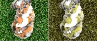
The medical term “third eyelid adenoma in dogs” is understandable for veterinarians or people who have already encountered this problem. For an ordinary person, it can be called anything, but it looks like loss or enlargement of the inner corner of the eye. At the same time, the eyeball itself remains unaffected, but if the adenoma of the third eyelid in a dog is not removed, this can lead to serious suffering for the animal.
Initially, you can observe the inside of the dog’s eyelid, as if the skin is pulled back and slightly sagging; this is the first symptom, in which it is better to immediately contact a veterinarian, which will allow you to quickly fix the problem and do it as cheaply as possible.
Third eyelid: where it is located and functions
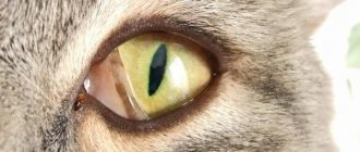
The third eyelid is a crescent fold located at the inner corner of the eye. It performs protective and auxiliary functions.
When you touch the eyelid and if you tilt your head, protection is immediately triggered - the cornea is covered by the third eyelid. This prevents all kinds of damage.
In the thickness of the third eyelid there is a lacrimal gland that produces about a third of all tear fluid. Also on its surface there is lymphoid tissue, which consists of numerous follicles. They are the most powerful immune defense for the eye.
Symptoms and first signs of the disease
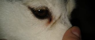
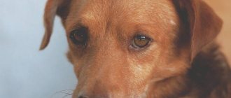
The first signs of the disease are the appearance of a large, bright pink neoplasm inside one or both eyes. In addition, the sick animal has the following symptoms:
- dry eyes;
- redness and swelling of the conjunctiva;
- swelling and redness of the eyelids;
- squinting of the eye;
- the appearance of mucous or mucopurulent discharge.
The disease occurs unexpectedly without any warning signs. Sometimes the loss of the nictitating membrane is preceded by the appearance of pasty gray-yellow masses in the corners of the eyes, indicating damage to the lacrimal glands.
Adenoma: risk groups
Pathology refers to prolapse of the lacrimal gland of the third eyelid. It goes beyond natural limits and becomes clearly visible to the eye.
Most often, such pathological changes in dogs are associated with age. There are also a number of breeds susceptible to this disease.
At what age does it most often occur?
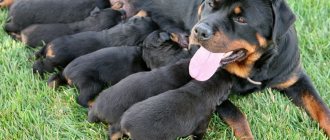
Animals under one year of age are susceptible to the disease. This is due to the fact that by this time the puppies’ ligaments have not yet had time to strengthen. Over time, they become more elastic and stronger, so the risk of developing pathology is significantly reduced.
Dog breeds susceptible to change
The development of the disease is often observed in breeds such as:
pugs;- Shar-Pei;
- Cane Corso;
- Yorkshire Terriers;
- Chinese Crested;
- cocker spaniels;
- chihuahua;
- bulldogs;
- Dobermans;
- Great Danes;
- German Shepherds.
Adenoma of the third century
The diagnosis of third eyelid adenoma in a dog is considered one of the most controversial in veterinary practice. In general, an adenoma is a benign tumor. Such a conclusion can be made only after a biopsy, that is, after taking the material obtained during the operation. The symptoms on the basis of which such a diagnosis is given, namely the expansion of the third eyelid and its bulging in the corner of the eye, are also characteristic of simple inflammation or hyperplasia.
It is important to understand that third eyelid adenomas are extremely rare in dogs. A diagnosis based only on an external examination can rightfully be considered unfounded by the owner. Especially when it comes to the pathology of the third eyelid in a puppy or 2-3 year old dog. Such a benign tumor is typical for animals that have crossed the threshold of 7 years or more.
If the pet is indeed diagnosed with this based on a biopsy, then treatment will necessarily include surgery. The specialist will differentiate the border between healthy and diseased tissue, determine the volume of the tumor, after which its complete removal will simply follow. Surgery is performed under general anesthesia, as it is quite painful. There are no other options for eliminating such an adenoma. However, there are some cases where the animal does not necessarily need to put itself at risk of unsuccessful surgery, here they are:
- the animal does not express concern that the third eyelid is swollen, does not rub its eyes with its paws, and the size of the swelling does not exceed a quarter of the size of the eye, that is, the dog sees perfectly;
- small size of adenoma;
- Over time, the tumor does not increase in size.
The decision about the operation is made by the owner. It is important to understand that the risk of third eyelid adenoma in dogs is minimal. In most cases, this is simply inflammation or volvulus, the treatment of which is much simpler.
Factors contributing to the development of the disease
Main reasons:
weakness of the ligaments designed to hold the gland in the correct position;- incorrect position of the eyelid cartilage;
- hyperplasia, which is a complication of previous leukemia;
- hereditary predisposition;
- damage to the eyelid during scratching, fighting or playing;
- diseases that provoke increased secretion of the gland.
Treatment
Therapeutic measures should be aimed at relieving inflammatory processes. However, there is no way to get rid of the pathology with medications.
Surgical intervention can be performed in two directions.
The first option is carried out by cutting, removing, excision of the gland. However, in this case, the eye loses the organ that produces the secretion and often such interference leads to dry keratitis.
Another solution is to preserve the lacrimal gland. In this case, an operation is performed, which aims to create a bed for the gland, place it in the anatomical space and fix it.
Surgery is performed under general anesthesia. There are no restrictions on the age of the animal.
During the operation, a pocket is formed from the conjunctiva, the gland is placed there and fixed with collagen threads. The suture is used with a submersible, thin thread and does not cause any disturbance to the animal. The suture material gradually dissolves and the sutures are not removed. Some clinics use monofilament atraumatic threads, which are removed 12-14 days after surgery under local anesthesia. The thread does not cause discomfort while “wearing” and the healing process is successful.
After the operation, the lacrimal gland is in the anatomically correct position and is not visible, but the third eyelid will still be swollen and slightly enlarged for some period.
Postoperative care requires the use of antibiotic drops. The drug “Sofradex” may be applicable, which has anti-inflammatory, bactericidal properties and prevents scratching, which allows you to avoid postoperative consequences.
Types of neoplasm
There are several types of adenomas:
- Cystic. Its structure is bag-like, closed.
Papillary. With this form of pathology, growths appear, which are papillae protruding into the lumen of the gland.- Polypoid. This is a kind of polyp formed as a result of hyperplasia.
Adenomas are of solid and tubular type. The first form is characterized by weak development of the stroma and fusion of the glandular epithelium into a single whole. In the second type of adenoma, narrow tubules are lined with epithelial cells.
Depending on the number of formations, the disease is divided into single and multiple. In the first case, only one node appears. With adenomatosis there are many of them.
Diseases of the third century
With the insanely fast pace of life, a person sometimes does not notice some of the changes that occur to his pet and lead to various diseases. This does not happen with diseases of the third eyelid; probably the very first place the owner will look is the eyes. Even minor changes will be noticeable immediately. Therefore, we will consider the most common diseases in dogs.
Volvulus
Occurs in young dogs up to one year old. This is due to the fact that in an unformed body, cartilage sometimes grows faster than the rest of the body, so the edge of the cartilage pushes out the third eyelid, and an incorrect anatomical position of the cartilage is observed towards the bend. There is a disruption in the normal functioning of all tissues of the eyelid, which leads to swelling, redness and hyperplasia (increase in the number of cells).
The most common problems are Newfoundland, Central Asian Shepherd, and Great Dane.
Treatment is carried out using surgery, excision of the cartilage occurs. Healthy, undamaged cartilage is not subject to excision.
Inflammation
As a rule, inflammation is caused by the action of pathogenic and conditionally pathogenic microorganisms, but in addition to this, there are a number of factors that cause this disease:
- damage, loss of integrity, eye injuries;
- getting small objects and dust into the eyes;
- irritation to smoke, various chemicals;
- with prolonged and uncontrolled administration of potent drugs;
- after a number of diseases, for example carnivore plague.
The owner can easily determine the signs of the inflammatory process. The dog has a slight prolapse of the eyelid, it is very hyperemic and has swelling, the size varies from a millimeter to a centimeter. The animal feels pain when touched near the eyelid, often lies down and tries to hide from bright light.
Treatment: use anti-inflammatory drugs, antiseptics. If the disease manifests itself as a result of infection, treatment is carried out with antibiotics and antiviral drugs.
Adenoma (true and false)
This is a common ophthalmological disease, which is characterized by a benign tumor neoplasm.
It is often confused with hyperplasia due to similar clinical signs - the organ enlarges and extends beyond its normal anatomical location, this is a false adenoma.
A true adenoma is confirmed by biopsy; if it confirms the presence of a tumor, then surgical treatment is used. The operation is determined by the doctor. If the tumor is small, does not interfere with the animal and does not limit the function of the eye, then surgical intervention is not required.
Hunting and decorative breeds are more susceptible to adenoma.
Hyperplasia
Hyperplasia is a pathological growth of tissue of the third eyelid. The clinical picture is very similar to an adenoma, only in this case the tissue proliferation is not caused by tumor processes. The nictitating membrane is enlarged, the dog’s third eyelid is red and falls out a little. The causes of hyperplasia have not been fully established. In order to treat hyperplasia, you need to know its etiology; treatment is generally conservative, but in some cases surgical intervention is resorted to.
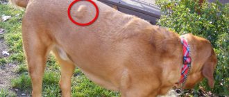
Loss (prolapse)
A characteristic sign is the third eyelid closing some part of the eye (half, a third of the eye). The cause of prolapse is often a weakening of the thin ligament that holds the third eyelid. Hair loss is not always an independent disease; sometimes it is a consequence of serious illnesses. This pathology can develop in the following cases:
- foreign body;
- eye injury;
- entropion of the eyelid;
- inflammatory processes or malignant/benign neoplasms of the bones of the upper jaw, nose, eye orbits.
Loss may occur in stressful situations (especially in small breeds), hidden infections, or parasitic diseases. As well as dogs suffering from ectropion of the eyelid (eversion of the eyelids).
Symptoms of the disease
When a tumor develops in a dog, characteristic symptoms appear that make it possible to distinguish an adenoma from other pathologies.
If the eye is affected, a small ball appears in the corner of the eyelid. The intensity of the shades may vary:
light red;- pinkish;
- red.
An enlarged gland leads to deterioration of vision and causes discomfort. Every time your pet moves their eyelid, the top layer of the eyeball is damaged. The cornea begins to itch very much.
The animal scratches the affected area, violating the integrity of the tissue structures, and the infection penetrates inside. There is a risk of developing follicular hypertrophy. Excessive lacrimation and discharge of pus cannot be ruled out.
At first, only one eye is affected. If treatment is not started in a timely manner, then after 1–1.5 months the pathological process will spread to the second organ of vision.
Disease prevention
Carrying out preventive measures will protect the animal from possible disease. Diseases of the third eyelid most often occur as a result of mechanical damage. Which means all kinds of animal injury prevention. This is especially true for hunting and guard dogs.
For owners when buying a dog, the main thing is to learn about all sorts of breed characteristics. Some dogs are more genetically predisposed to third eyelid disease. If various pathologies and symptoms occur, you need to contact a specialist. Doctors warn about the possible consequences of home treatment. Disease of the third eyelid in a dog requires attention and treatment in the clinic. During treatment, the owner must remain close to the pet at all times to prevent the dog from scratching the eye and getting an infection into the wound. During treatment, you should also reduce or eliminate walks outside (especially in the cold season).
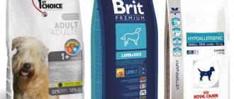
During treatment, you also need to monitor your diet. Doctors recommend using dietary food to avoid allergic reactions and complications. Additionally, after treatment, immunostimulants and vitamin supplements should be used, which should be aimed at improving the microflora after taking antibiotics. Therefore, avoiding walks and proper treatment and care can speed up the animal’s recovery process. Every owner should remember that eye diseases in dogs can lead to blindness or death if not treated promptly.
Currently reading:
- Neoplasms and infections in diseases of the eyes of dogs
- Thyroid dysfunction in dogs (hypothyroidism)
- Blindness is a faithful companion of entropion in a dog
- Fear of blindness due to Blepharitis in a dog
Treatment of adenoma
Before determining treatment tactics, the veterinarian conducts a comprehensive diagnosis. It includes a number of activities:
Biopsy. With its help, it is possible to find out whether a formation is benign or malignant.- Blood chemistry.
- Suctioning fluid from lymph nodes.
Treatment begins only after confirmation of the diagnosis.
Reduction and suturing
Previously, veterinarians were convinced that only removal of affected tissue was effective. Now doctors can perform treatment even without surgery.
Conservative methods are used. The modified gland is reduced to its natural position and sutured. But this method of therapy is allowed only if an adenoma is detected at an early stage of its development. In advanced forms of pathology, such actions are ineffective.
If the dog breeder contacts the veterinary clinic in a timely manner, repositioning the gland does not cause any particular difficulties. The procedure is carried out using ordinary tweezers. To prevent the gland from falling out again, it is sutured immediately afterwards.
Surgical intervention
To completely eliminate inflammatory processes in the area of the glandular epithelium, they resort to surgery. Before this, therapy is carried out using antibiotics. Antimicrobial drugs with a wide spectrum of action are prescribed.
The duration of antibiotic use directly depends on the individual characteristics of the pet and its health status.
The course of antibiotic therapy lasts from several days to one week. After this, an operation is performed.
Progress of surgical intervention:
The animal is fixed on the couch and placed on its side.- A painkiller is administered.
- The third eyelid is grabbed with a clamp and pulled to the maximum distance from the palpebral fissure.
- Curved scissors are used to remove the eyelid.
- Stitches are placed on the wound.
- The affected tissues are treated with an antiseptic.
Surgery to eliminate the disease
Treatment methods for this pathology have been controversial among doctors for many years. Ten years ago they came to a consensus that there is one way - this is surgical intervention, removal of the prolapsed gland. But it was soon discovered that the dog then developed dry kerato-conjunctivitis, which progressed for a long time and disappeared only after two years. The gland also produces approximately 40% of tears.
Therefore, they put forward a decision that the gland should be returned to its place so that it continues to work normally - to produce tears, which have the function of protecting the eyeball. Surgeons must prevent the eye and gland from drying out and becoming inflamed.
If the pathology has not reached a serious stage, it is possible to realign the gland by a specialist using ordinary tweezers. This option is not considered effective, since there is a high probability of repeated loss. The procedure is carried out if no more than a day has passed.
Surgical reduction of the gland remains the current method. If it is severely inflamed, treatment with antibiotics will be required before surgery. Therapy can last several days.
The dog's affected eye is instilled with a 0.25% solution of Levomycetin. The procedure is carried out once a day. Experts advise laying a special film with Kanamycin. At the same time, 1-2 drops of a 1% Dicaine solution are instilled.
There are two options for surgical reduction: the “pocket” technique and its modifications and fixation of the gland to different orbital structures using non-absorbable sutures and their modifications. The first option is the most common. The operation is carried out quite simply without special equipment.
Video: surgical treatment of third eyelid adenoma in dogs
After immersing the pet in anesthesia, applying antiseptics, and fixing the eyelid speculum, the eyelid is secured by the edge with clamps. Then it is pulled out of the conjunctival sac so that the bulbar region of the conjunctiva and the prolapsed gland are turned towards the doctor. Incisions are made under and above the lacrimal gland, maintaining a distance between them.
The specialist immerses the lacrimal gland deep into the orbit and closes the conjunctiva in the incision areas. Their edges are sutured using absorbable sutures, the lacrimal gland remains in the pocket, preventing its appearance on the surface. At the end of the surgery, the dog requires a week of simple care.
Removal of the third eyelid is necessary only in particularly advanced cases - with degenerative or necrotic changes. During the operation, general anesthesia is used. There is no need for deep anesthesia.
The process involves a microscope and specific ophthalmons. The specialist uses them to “package” education. This prevents injury and the appearance of rough scars. The procedure usually lasts a quarter of an hour. It is allowed for dogs of any age, but the consequences for older pets can be unpredictable. The price of the operation depends on the size of the animal, the region and the specific clinic.
Recommendations for caring for your pet after surgery
Sometimes after surgery, pets are left under the supervision of a doctor. To relieve swelling, antimicrobial drugs and antibiotics are prescribed. If the procedure is carried out correctly, no relapse is observed. Great Danes are an exception; they often develop new formations.
After surgery, the owner must provide the dog with proper care at home. A special protective collar will be required. It is used for no more than 15 days. It is recommended to use eye drops and ointments. With proper treatment, signs of the disease disappear within a month.
Caring for the animal after surgery
During the postoperative period, the dog owner needs to provide the pet with proper care. Thanks to this, rehabilitation will be quick and will not cause complications.
Drug treatment
After the operation, the veterinarian prescribes medications to eliminate swelling.
For this purpose, antibiotics and antiseptics are used. You cannot choose treatment on your own. Treatment tactics and medications should be agreed with your doctor.
Immobilization collar
To prevent the dog from damaging the operated organ, care must be taken to have an immobilizing collar.
The animal will need to wear it for 2-4 weeks. The device is necessary to prevent scratching of the eye and penetration of infection into the affected tissue.
How to help a dog after tumor removal
After the operation, there is a risk of decreased performance of the organ of vision. The animal may experience dry eye syndrome, caused by insufficient tear production.
The dog owner needs to use special means to replace tear fluid. They come in the form of drops.
Non-surgical treatment methods
Specialists do not resort to surgery if the animal has absolute contraindications to the use of anesthesia or if the owner does not want to treat the disease surgically.
If there is no oncological nature of the pathology, the neoplasm is small in size, which does not cause discomfort to the animal, a non-surgical method of treating third eyelid adenoma is justified.
Treatment consists of manual reduction followed by antibacterial and hormonal eye drops.
Interesting! Non-surgical methods, unfortunately, are ineffective; in 80% of cases, conservative therapy leads to relapses and complications.
Prevention of adenoma development
To reduce the risk of developing adenoma, you need to adhere to the following recommendations:
- If you identify any health problems in your dog, immediately seek help from a veterinarian;
- do not allow your pet to scratch its eyes, this will prevent infection;
undergo regular examination by a veterinary ophthalmologist;- do not self-medicate in case of mechanical damage to the organ of vision;
- treat the eyes with antibacterial agents, after consulting with a veterinarian;
- When taking medications, give the dog vitamin-mineral complexes.
Prevention and care for dog eyes
Eye pathologies in animals can be prevented by following simple preventive measures. Dog eye care consists of the following regular activities:
- daily examination of the animal’s organs of vision;
- rubbing the pet's eyes with special lotions in the direction from the outer corner of the eye to the nose;
- when washing the animal, avoid getting water and shampoo into the dog’s eyes;
- trimming long hair on the face;
- If there is redness, swelling, discharge, spots or growths in the eyes, immediately contact a specialist.
Forms of pathology
The third eyelid can only be visually seen when the dog blinks. Due to the fact that the lacrimal glands are also located at the base of the semilunar fold, it also performs the function of moisturizing the outer membrane of the eye. Lymphoid tissue serves as an additional barrier against infections.
And if in a normal state this membrane is practically invisible, then when inflamed it turns red, swells, bulges outward and noticeably increases in size. There are three main forms of pathology:
Treatment of the third eyelid in dogs
A detailed examination of the dog’s third eyelid, identifying the specific causes of the disorder, allows us to draw up a specific treatment plan for the animal. Usually it is based on traditional drug therapy, but in complex cases the help of a surgeon is simply necessary.
With the help of drugs
Traditional therapy involves the use of antispasmodics and antiseptics, in addition to which painkillers and corticosteroids are sometimes prescribed. Using antiseptics, the affected eye is washed, thereby preventing the development of infectious processes in it, anti-inflammatory medications prevent suppuration of damaged areas of the third eyelid, and corticosteroids regulate metabolic processes in the body and contribute to a faster recovery of the animal. There are many examples of suitable medications, but among the most accessible and effective topical medications, tetracycline ointment is usually distinguished.
Important! The duration of the course of therapy and the dosage of the selected drug for each dog are calculated individually, taking into account the age and weight of the animal, as well as the presence of chronic ailments, which in some situations may become a contraindication to treatment with a certain composition.
Removal operation
A surgical solution to problems with the third eyelid is relevant in case of mechanical damage, loss or excessive growth of tissue (for example, hyperplasia), with a possible change in their normal structure and the appearance of malignant neoplasms. Complete removal of the eyelid (especially with the lacrimal gland) is an extreme measure and in practice is used in isolated cases. If the operation to fix the lacrimal gland to the zygomatic bone is successful, the integrity and mobility of the film can be preserved, and recurrent eyelid loss is eliminated by almost 90%.
The recovery period takes 10–15 days and until the eye heals well, it is advisable to put a special collar on the dog. It will prevent scratching and infection from entering the tissue. In some cases, before the operation, the pet is prescribed preparatory treatment, using a course of antibiotics. The main goal of this preparation is to reduce the likelihood of complications during surgery.