Causes of peripheral edema
Increased permeability of the vascular wall caused by the action of inflammatory mediators and bacterial toxins on it.
Inflammatory mediators are special substances secreted from special cells called mast cells, or basophils. Such swelling occurs around a traumatic skin lesion, or other inflammatory focus, and is an integral component of inflammation. The body's generalized response to infection, or sepsis, is also accompanied by a massive release of inflammatory mediators and can lead to generalized edema. Local swelling of the face and neck, along with redness of the skin and mucous membranes, can accompany acutely developing allergic reactions. A similar thing can happen with an insect bite - wasp, bee, horsefly.
Fluid can penetrate more through the vascular wall when hydrostatic pressure in the vessels increases, which occurs due to fluid and sodium retention in the body or due to slower blood flow in the vessels. This happens quite rarely in severe congestive heart failure or renal failure.
The fluid located in the interstitium, between the cells, is also in motion, and flows through the lymphatic vessels, then flowing into the veins. Obstructions that impair lymphatic drainage can also lead to peripheral edema. For example, this happens with inflammation of the lymphatic vessels (lymphadenitis), their compression, removal of lymph nodes during oncological operations.
If a tumor is found in a dog’s neck under the jaw, treatment directly depends on its cause. Inflammatory formations on the pet's neck can be successfully treated with the use of antibacterial drugs. In some cases, a veterinarian resorts to opening the abscess.
Attention should also be paid to the treatment of warts and papillomas. As a rule, professionals recommend removing these tumors. Firstly, the growths can be injured when the pet walks and rests, and secondly, one should not discount the possibility of such lumps degenerating into malignant structures.
With the help of a surgical scalpel it is possible to stop a cancerous tumor in a dog. Modern veterinary medicine significantly prolongs life and improves its quality when a terrible diagnosis is established. To treat a dog, laser methods are used to target the tumor, as well as chemotherapy.
A tumor on the neck of a four-legged family member requires the owner not to delay a visit to a specialized institution. A veterinary specialist will determine the nature of the neoplasm and determine a therapeutic strategy. In some cases, a furry patient will need surgery.
There are many reasons why a tumor appears. It happens that they can be seen on one and the other half of the muzzle. This may develop due to:
- Infections/cellulitis;
- Allergies whose causes are difficult to determine;
- Poisoning with paracetamol or products containing it;
- Collections of liquid or clotted blood;
- Inflammation of muscle tissue;
- Inflammation of skeletal muscles;
Diagnosis
A tumor in a dog can be the result of an allergic reaction after an insect bite or taking paracetamol. The site of the tumor causes pain when palpated. As a rule, this is accompanied by elevated temperature and poor health of the animal. The formation grows to the tissue, but there are exceptions to the general rules.
The tumor develops as a hematoma after injury. In this case, only one side of the animal's face swells. The tumor, when pressed, can cause pain, and in some animals the area may even turn blue.
Chondrogenic sarcoma, osteogenic sarcoma, lymphoid sarcoma and fibroid sarcoma are types of malignant tumors in dogs that can appear on the dog's face. Swelling of the lymph nodes located under the jaw can also appear as a tumor on the face. It can cause pain and appear as an open wound. Swelling is located in the area of the lower facial bone or in the upper area of the head.
By palpation, it is important to check the tumor for mobility, painlessness during examination, and the intensity of development. Attention is paid to the well-being of the animal. If you notice the first symptoms of formations, call your doctor for a diagnosis and prescribe a course of treatment.
Types of bumps on the neck
The tumor can develop in connection with purulent inflammation of the cervical tissues. Abscesses are localized in two layers: subcutaneous and muscular. The formations occur as a result of an insect bite or other allergic reaction. The tumor may be a consequence of the papilloma virus. Dark growths appear on the skin, they are painless and do not bother the animal. The cause of the virus is unknown.
The tumor may be the result of a pyogenic coccus. Pus begins to accumulate in the animal's neck area. At the initial stage, the outer layers are damaged. If you do not contact a veterinarian at this stage, the pus can affect the subcutaneous tissue and muscles.
Tumors can cause benign or malignant neoplasms. From the very beginning, their size is insignificant and it cannot harm the dog or limit his active life. If you are going to visit a doctor, it is advisable not to feed or water your pet for several hours, as he may prescribe a number of diagnostic procedures for you.
Tumors in the throat can cause a rupture of the trachea, so if the dog's neck looks several times larger than its usual size. The animal has difficulty breathing. His gums have a purple or blue tint and the symptoms do not go away after a few hours, go to the doctor immediately.
Throat swelling in an animal often occurs simultaneously with swelling of the face. Symptoms may include difficulty eating, metrorrhagia, and a rotten odor. The animal's eyes may become swollen or swollen.
Formations in the throat area that arise as a result of uncontrolled cell proliferation. They need treatment. And this does not depend on whether they are malignant or not. Surgery to remove the swelling or radiation therapy will prove to be an effective treatment method.
According to a number of statistics, this type of tumor makes up more than half of all cancers in dogs.
Mostly female dogs are affected by the disease, but only in one percent of cases is it diagnosed in males. Most often, breast formations appear at the age of ten, sometimes this can happen earlier. The disease is extremely rare in animals under four years of age.
The tumor is described as an uncharacteristic growth of iron tissue that cannot be controlled by the body. It contains atypical cells with an irregular structure. They differ from functional cells, both in their operating principle and in structure.
Cells with a different nature begin to quickly divide and transport their impulses throughout the genetic code. Tumor tissue grows rapidly by cell division. The process of how exactly a tumor arises and develops has been studied for a long time, but it is impossible to obtain more complete information about the process. This is due to difficulties in conducting research.
A malignant formation can develop up to several years without clinically manifesting itself. How quickly a specific tumor will grow and develop is personal in any case, so the prognosis for the animal should always be approximate from the very beginning.
Neoplasms on a dog's chest are divided into benign and malignant. Among all tumors, more than half of them belong to the latter type. The most common is cancer.
A feature of malignant tumors is their metastasis; they can spread throughout the body through the bloodstream or lymph. Harmful cells that have spread throughout the body, in some cases, cause tumors to appear in other organs.
Cancer cells often cause the formation of secondary growth foci in the lymphatic system, and only then do they develop in the circulatory system. Therefore, the first signal of disease progression is the appearance of a secondary tumor in the closest lymph node.
For distant metastasis, the most likely area of spread of lesions will be the respiratory system, but growth in other systems is also likely. The fact that tumors are benign and also malignant is conditional, because any benign manifestation can, as a result of exposure to a carcinogen, develop into cancer.
Tumors in adult dogs or puppies can also occur in the abdominal cavity. They are often found in the area of the spleen. The formation, having reached a certain size, with chaotic movement during a strong impact, can become deformed or rupture. As a result, bleeding occurs, and ultimately death; sometimes the animal does not even have time to be delivered to the clinic.
The owners become aware of the tumors when they reach a critical size and begin to become an obstacle to the normal functionality of the body. If measures are not taken, the pet’s condition will worsen, and surgical intervention is inevitable.
Treatment of tumors in dogs, causes and diagnosis
Every year, various dog diseases appear that affect the quality of life of our pets. Today we will talk about cancers ( canine tumors ) that can occur in dogs. We will try to consider the main causes of its appearance , characteristic symptoms and possible methods of treating tumors in dogs .
Tumors in dogs: lymphoma
is a cancer of the lymphatic tissue in a dog's body . This type of tumor accounts for about 20% of the total number of calls for suspected cancer:
The causes of lymphoma - there is no clear answer to the reasons why lymphoma appears in a dog .
But scientists identify some factors that may affect its presence.
These include the age characteristics of the dog, the state of immunity and the influence of other diseases against which lymphoma appears. A breed prone to cancer - Labrador
;
symptoms of lymphoma - characteristic signs for this disease are: enlarged lymph nodes, especially in the jaw area, near the shoulder blades and knee. Possible swelling of the tonsils, general lethargy and apathy. If the lymphatic tissue of internal organs is damaged - diarrhea, abdominal pain, shortness of breath;
Treatment of lymphoma is possible with either a single steroid-based drug or complex chemotherapy.
Tumors in dogs: hemangiosarcoma
Hemangiosarcoma - this tumor develops from the cells that line the blood vessels.
causes of hemangiosarcoma - these include both the consequences of diseases and genetic predisposition. The breeds that are most prone to hemangiosarcoma are Labradors and German Shepherds;
symptoms of hemangiosarcoma - the great danger of this disease is that for some time it can be asymptomatic and not cause visible harm to the dog.
As a rule, the appearance of the disease at an early stage is recorded when the animal is examined by a specialist during a routine visit, or in the presence of obvious symptoms in advanced stages.
Such signs are weakness, shortness of breath, apathy, pale gums;
Treatment of hemangiosarcoma - treatment of tumors in dogs depends on many factors. It is very important to notice the disease in the early stages, this will help you complete the course of treatment successfully.
In the later stages, even an enhanced chemotherapy regimen may not help and the animal will die.
Treatment is carried out through surgery, where the bleeding is stopped and the affected organ (mainly the spleen) is removed, after which a course of chemotherapy is prescribed.
Osteogenic sarcoma - tumors in dogs
Osteogenic sarcoma is a type of tumor that affects the bone tissue of a pet.
causes of osteogenic sarcoma - the main factors influencing the occurrence of the disease include the genetic characteristics of breeds. Great Danes and similar animals with massive bone skeletons are at risk. They are 200% more susceptible to the disease than representatives of small breeds (Chihuahua, toy terrier, etc.);
symptoms of osteogenic sarcoma - weakness, gait disturbance, clumsiness, apathy;
treatment of osteogenic sarcoma - in order to completely eliminate the pathology of bone tissue, an operation to amputate the affected limb is usually prescribed. After surgery, a course of chemotherapy is required. If you refuse surgery, treatment with medications alone can prolong your pet's life by only 6-12 months.
Mastocytoma - tumors in dogs
Mastocytoma - in the animal’s body there are mast cells that are responsible for the state of the immune system and allergic reactions. Mastiocytoma is a neoplasm that consists of these cells and appears on the skin of an animal.
The causes of mastiocytoma are the main factor; it is a genetic predisposition. Much also depends on the breed. Bulldogs and boxers are at risk of developing mastiocytoma;
symptoms of mastiocytoma - you can visually notice changes on the dog’s skin. Growths and formations may appear. The general condition may be unchanged;
treatment of mastiocytoma - treatment methods depend on where the formation is located. In some cases I prescribe only surgery, in others only chemotherapy, and sometimes complex treatment is necessary.
Melanoma - tumors in dogs
Melanoma is a tumor of skin tissue, specifically the pigmented cells of the skin.
causes of melanoma - since the main element of the lesion is dark skin cells, dogs of certain breeds are mainly susceptible to infection. These breeds include Doberman, Toy Terrier and the like. The formation can appear anywhere, since skin cells are found throughout the body;
symptoms of melanoma - lack of appetite, deterioration in the dog’s general condition, noticeable abnormalities in the oral cavity;
Treatment of melanoma should be prescribed by the attending physician. Treatment methods are determined based on the histology results obtained. This mainly involves surgery, chemotherapy or radiation therapy. Removing a tumor from a dog requires consultation with other specialists.
Squamous cell carcinoma - tumors in dogs
Squamous cell carcinoma - this type of formation is characterized by damage to tissues located on the skin and in the mouth of the animal.
causes of squamous cell carcinoma - these include various reactions in the animal’s body. External factors can also influence the development of the disease;
symptoms of squamous cell carcinoma - since this type of cancer can affect the cells of the oral cavity, the dog may experience loss of appetite due to pain, as well as lethargy, weakness, apathy;
treatment of squamous cell carcinoma - if a growth is found on the skin in most cases, surgery to remove the tumor takes place without consequences and completely helps to get rid of the disease.
If the formation is in the mouth, difficulties may arise. They are associated with the difficulty of identifying oncology until the disease progresses to the last stage.
If the disease is detected late, the animal lives for about a year, after which it dies.
Breast cancer - tumors in dogs
Breast cancer is a very common cancer seen in dogs. The tumor appears in the mammary glands and damages nearby tissues.
causes of breast cancer - hormonal changes in the body can provoke breast cancer. They can be caused by the use of hormonal drugs to suppress animal urges during estrus. Also, if sterilization is not carried out in a timely manner, the risk of the disease increases;
symptoms of breast cancer - characteristic signs include the presence of seals in the nipple area, the animal can constantly lick the site of the tumor, itching, weakness and apathy;
Treatment of breast cancer - involves the removal of a breast (one or a whole series) through surgery. Chemotherapy is also prescribed, which is necessary in cases of disease progression to other parts of the body. It is important to start treatment on time to avoid complications.
Anal gland cancer - tumors in dogs
Anal gland cancer - in dogs, in the area of the anus there are two sacs with glands that help the intestines or excretion pathways function normally. This type of tumor affects these glands.
Causes of cancer of the paraanal glands - the reasons may be different. Sometimes the presence of cancer is diagnosed when blood counts change and calcium levels are elevated. Poor nutrition and intestinal pathologies can also be a cause. Obesity and weak muscle tone of the anus and rectum can become determining factors in oncology;
symptoms of cancer of the paraanal glands - a clear sign of this disease can be a change in the dog’s behavior. It is expressed in the appearance of the habit of “riding on your butt on the floor.”
The animal literally sits on its fifth point and begins to actively move. There may be redness at the anus, as well as suppuration or swelling.
All this is accompanied by the pet’s irritability, reluctance to move actively, loss of appetite, problems with bowel movements;
treatment of paraanal gland cancer - when a paraanal tumor is diagnosed in dogs, the doctor prescribes surgery to remove the tumor that has appeared. After surgery, radiation therapy may be performed. If the disease begins to spread to other tissues, chemotherapy is used.
Transitional cell carcinoma - tumors in dogs
Transitional cell carcinoma in dogs affects the organs of the urinary system.
causes of transitional cell carcinoma - genetic predisposition can cause cancer, as well as the consequences of various diseases that affect the organs of the genitourinary system. Although this type of tumor is considered local, it can progress and spread to other areas of the body;
symptoms of transitional cell carcinoma - symptoms depend on the location of the tumor. Generally, there may be blood in the urine and problems with urination;
Treatment of transitional cell cancer involves surgery and a course of chemotherapy.
Soft tissue sarcoma
Sarcomas are a group of different types of tumors that affect the soft tissue on your pet's body.
the causes of sarcoma are various chemical processes in the animal’s body, genetic predisposition, breed characteristics and the consequences of various diseases;
Symptoms of sarcoma are lumps in different parts of the body. Weakness of the pet, apathy and decreased physical activity;
Treatment of sarcoma - involves surgical removal of tumors, possibly a course of chemotherapy. Before carrying out therapeutic measures, it is necessary to conduct a competent diagnosis of the disease in order to make an accurate diagnosis.
All described characteristics of tumors in dogs are purely informative. We do not encourage you to self-diagnose diseases, much less self-medicate.
Any amateur activity can cause harm to your pet, which will affect its health. Providing assistance without certain knowledge and skills can lead to death for the dog.
To avoid such situations, seek help only from qualified veterinary centers.
Tumors in dogs - conclusion
We looked at the different types of tumors that can occur in dogs. All neoplasms have different etiologies and symptoms.
Knowing the signs and causes of tumors in dogs, owners have the opportunity to quickly respond to manifestations of the disease and seek help as quickly as possible. This will help protect the animal from complications arising during the course of the disease.
Oncological diseases progress very quickly, so catching cancer at an early stage is the main task of the owner.
To avoid becoming a victim of scammers and amateurs, you must contact only specialized veterinary centers
.
Ours offers the services of only highly qualified specialists who meet all the requirements of veterinary professionals.
The equipment used for examinations and procedures meets European quality standards, and the drugs used are certified.
The range of services we provide is very diverse. We advise you to pay attention to. It will help save your time, and also relieve the animal from stress, which may occur during transportation. When talking about cancer, it is worth remembering that stress is the worst enemy that contributes to the development of the disease.
We are always happy to help you. Take care of your pets!
Source: https://ya-vet.com/onkolog/lechenie-opuholej-u-sobak
Mechanical damage to blood vessels
Just like in humans, dogs' blood vessels lose elasticity over time. Even with a minor impact, capillaries and small veins burst: blood flows out and fills the free space. A compaction forms on the surface.
It will not be difficult for an attentive owner to determine the nature of the formation: at the moment of rupture, the dog experiences pain, he worries and whines. Then he calms down, but upon inspection he tries to dodge. The dog's behavior, activity and appetite do not change.
This lump is initially hard to the touch, then softens and completely resolves. To alleviate your pet’s condition, it is recommended to apply an ice compress for 10-15 minutes immediately after detection. To do this, wrap a plastic container with ice in a dry cloth and press it to your neck. It is not recommended to keep the lotion longer: instead of benefiting, the cold will cause harm.
Lump on a dog's neck after an injection
When walking, pets do not sit still: they are in constant interaction with their relatives and surrounding objects. The owner is not always able to assess the degree of safety of his pet. Therefore, the moment of defeat remains unnoticeable: the animal does not feel pain and continues to play happily. And some time after the injection, a lump appears on the dog’s neck.
First, you need to carefully feel your pet: perhaps there is a splinter left in the wound. It needs to be carefully removed. Then apply and fix the tampon with Vishnevsky ointment for 12 hours. Usually, these measures are enough for the dog to recover.
The animal will try to pull off the bandage, so you should put a special collar on it for the entire duration of treatment.
From the above it is clear that treatment of edema is, first of all, treatment of the underlying disease that caused the edema.
The causes of edema associated with local inflammation or injury are relatively easy to recognize. In this case, measures are taken to treat the cause of inflammation - most often antibiotics and antiseptics. Chronic joint damage (arthrosis), with significant stress, can also lead to inflammation and swelling of the tissues of the joint and paw.
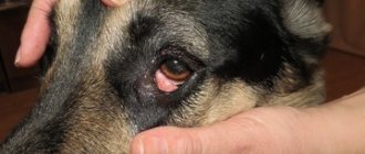
Swelling of the face or neck (Quincke's edema), which is a symptom of an allergy, can be life-threatening, in which case it is necessary to immediately bring the animal to the clinic. It is important to know that antihistamines, such as diphenhydramine, suprastin, and others, are rarely effective in dogs and cats, and their effectiveness cannot be predicted, therefore, first aid for angioedema is the restoration of airway patency, if it is impaired, adrenaline, because it inhibits the release of inflammatory mediators from mast cells, and steroid hormones (prednisolone or dexamethasone), because they inhibit allergic reactions at all stages of their development. Quincke's edema rarely develops on drugs given orally, mainly on drugs administered parenterally, that is, by injection.
The drug that caused the allergy is prohibited from being administered in the future - tell all doctors who will subsequently treat your animal about this.
Edema due to low blood albumin can be diagnosed by measuring albumin and total protein levels (blood chemistry test). Low albumin levels can be corrected by intravenous administration of human albumin and a nutritious diet. In addition, transfusion of colloidal solutions (Refortan, Infucol) and careful use of diuretics can help in the fight against peripheral edema.
Do not forget that this type of peripheral edema is also only a symptom, and if your animal does not have a clear reason for protein loss (malnutrition or diarrhea), it is necessary to examine the internal organs (liver, kidneys) - a biochemical blood test, ultrasound is also used for this , general urine analysis.
Peripheral edema due to chronic heart failure is quite rare, however, when accepting an animal with edema, the doctor must, at least on the basis of examination data (palpation of the pulse, auscultation of the heart, examination of the mucous membranes), form an opinion about the functioning of the heart.
Local edema due to impaired venous and lymphatic drainage is also quite rare. You may encounter this type of edema, for example, if an intravenous catheter has been in the animal’s paw for a long time - then you can loosen the plaster securing the catheter, or, after agreeing with your doctor, remove the catheter completely.
The diagnosis can be made based on examination and characteristic clinical signs alone. Moreover, from excitement at the sight of a veterinarian, the dog usually begins to wheeze even louder and more clearly. Examination of the larynx using a laryngoscope, endoscope or ultrasound is possible only with the preliminary administration of sedatives, since otherwise it will still not be possible to properly examine the swollen and irritated organ.
Benign formations under the skin:
- Hematomas. A soft formation occurs due to disruption of the integrity of blood vessels due to injury. In rare cases, subcutaneous hemorrhages may be painful to palpation.
- Lymphoextravasate. Severe trauma can lead to rupture of not only blood vessels, but also lymphatic ones, which is accompanied by the accumulation of lymph under the skin in the form of a lump.
- Abscess. A bite from a relative, a puncture wound, or a deep splinter are often complicated by a bacterial infection, resulting in swelling. Violation of the rules of asepsis and antiseptics during injections leads to the development of an inflammatory process.
- Insect bites (bees, wasps, horseflies).
- Warts, papillomas. Such bumps do not cause concern to the pet and are typical for short-haired individuals. As a rule, the growths have a brown color and a heterogeneous (lumpy) surface. Papillomas resemble cauliflower, soft and loose in consistency. The owner should be alert to a wart or papilloma from which blood is oozing.
- Purulent skin diseases (pyoderma). The bumps are caused by the development of a bacterial infection. Pyoderma bumps are found not only on the neck, but also quickly spread throughout the pet’s body.
- Cyst. A cystic formation on the neck of dogs is a rare occurrence and is formed when the ducts are blocked or compressed.
- Lipoma. A benign neoplasm under the skin is a ball-shaped compaction and consists of adipose and connective tissue. The lump usually occurs in dogs over 5 years of age.
- Hemangioma, keratoacanthoma, fibroma. Neoplastic tumors such as trichoepithelioma and pilomatrixoma are less commonly diagnosed. Typical for animals older than 5-6 years. In some cases, there is a breed predisposition.
Malignant tumors in dogs include primarily hemangiosarcoma, melanoma, liposarcoma, and fibrosarcoma. Mast cell tumors develop quickly and are characterized by infiltration into other tissues and the development of metastases. The general condition of the four-legged friend worsens. The dog loses its appetite, becomes lethargic and inactive.
Multicentric lymphoma
What to do if detected:
- it is necessary to examine the pet for pain and signs of inflammation;
- You should not self-medicate, but visit a specialized institution and find out the nature of the neoplasm.
Diagnosis of an animal includes:
- thorough examination of the animal, palpation
- biopsy - taking biological material followed by bacteriological or cytological examination;
- general and biochemical blood test;
- X-ray examination, computer and magnetic resonance imaging.
Treatment of a tumor on the neck in dogs:
- Inflammatory formations on the pet's neck can be successfully treated with the use of antibacterial drugs.
- In some cases, a veterinarian resorts to opening the abscess.
- Papillomas and warts are removed.
- Cancerous tumors are removed surgically; laser methods of treating the tumor and chemotherapy are also used.
Read more in our article about tumors on the neck in dogs, causes and treatment.
Read in this article
Types of bumps on the neck
The tumor may be a consequence of the development of a uterine infection or an inflammatory reaction in it. Every day, about ten operations are performed in the veterinary clinic to remove the uterus from an animal. According to the nature of the progression of the disease, it is divided into acute and chronic forms. The latter may take several years, but will definitely end with surgical removal of the organ.
Possible causes of edema in dogs
It should be remembered that swelling of the paws or other parts of the body is, first of all, a clinical sign, and not a separate disease. This clinical sign occurs due to excessive release of fluid from the vessels. Edema of the body itself has a certain classification. For example, swelling can be mild, pronounced, or simply pronounced. As for the prevalence of edema, they can be divided into local (located in a specific area) and generalized (over the entire body of the animal). Such swelling can be caused by a number of special factors and diseases, including:
- Mechanical damage to the skin;
- The appearance of a focus of inflammation;
- Infection or sepsis (these disorders usually provoke generalized edema);
- Various types of allergies (this reaction usually affects the dog's face and neck);
- Insect bites, such as bees or wasps;
- A noticeable decrease in albumin in the dog’s blood, caused by a number of liver diseases, including cirrhosis, hepatitis, and tumors of this organ;
- Too much protein loss occurs due to the progression of diseases such as chronic or acute renal failure.
- Diseases of the gastrointestinal tract, accompanied by frequent diarrhea.
- An increase in pressure in the blood vessels, usually due to a slowdown in blood flow. This often happens with chronic renal or heart failure.
- Arthrosis.
- Inflammation of the lymph nodes or their removal through surgery.
Insect bites
Thick fur protects pets from insect bites: wasps, hornets, flies, bees. And instinct does not allow dogs to approach dangerous neighbors. But sometimes an attack does occur for various reasons:
- swarming of bees, wasps, hornets;
- hot weather leads to an increase in the horsefly population;
- seasonal activity of insects;
- moving to regions with unfamiliar insects.
If a dog is attacked, the bites are concentrated on the neck. When palpating, the finger comes across a hard lump on the dog’s neck. The animal's behavior changes: its temperature rises, and with several bites, the amount of poison entering the bloodstream can lead to the death of the dog.
Hornets and wasps do not leave stings, but if a bee attacks, the sting must be removed immediately. Then you need to lubricate the bite site with an alcohol-containing solution, and provide the animal with plenty of drink with an antihistamine (dissolve a suprastin tablet in water or drop syrup).
FAQ
Only angioedema requires immediate medical attention. If, when administering any drug, your animal's muzzle suddenly swells, the mucous membranes of the mouth become intensely red or, on the contrary, turn pale, vomiting and rapid, labored breathing, sometimes with wheezing, occur, immediately take the animal to the clinic.
Most likely no. Typical actions of the doctor at the appointment are as follows: if the animal is choking, it may be necessary to intubate it - insert a tube into the trachea through the mouth under anesthesia, or perform a tracheostomy. Give oxygen to breathe. Administer adrenaline 0.01 mg/kg intramuscularly, prednisolone 2 mg/kg intravenously or intramuscularly, and establish intravenous drip administration of fluid.
Other types of edema require observation and planned clarification of their causes by a therapist, because in themselves they do not threaten the animal in any way.
Maybe, but a healthy animal’s paws don’t just swell, even an untrained one and after a significant load. A routine examination is necessary to identify possible problems with joints or internal organs.
No, until you see a doctor and find out the cause of the swelling, avoid any treatment. You will not eliminate the cause of the swelling, and the doctor may then have to deal with the consequences of such “treatment.” For severe inflammation of the skin, a dermatologist, in addition to the main treatment, may recommend compresses with magnesium sulfate (magnesia), which relieve inflammatory swelling well, but wait with them before consulting a doctor.
If the bite does not affect the general condition of the animal and does not cause severe swelling, just watch it; you don’t need to do anything, except perhaps treating the bite site with an antiseptic (iodine or brilliant green). A bite that causes significant swelling, itching, or anxiety should be shown to a doctor. A single injection of corticosteroids will help quickly relieve unpleasant symptoms if they are significant. Quincke's edema or anaphylactic shock that occurs after a bite naturally requires immediate medical attention.
Diseases accompanied by edematous and congestive phenomena are far from uncommon in both medical and veterinary practice. Some of them, such as laryngeal edema in a dog, are not only extremely undesirable, but also fraught with serious consequences for the entire animal’s body.
As you might guess, this is the name for the process of swelling of the tissues of the larynx. This organ is very important, as it plays an important role in the process of breathing and sound communication. If the larynx is swollen, the respiratory function does not work well, the animal breathes heavily and hoarsely, cannot eat or drink, since the swallowing movement cannot be performed.
The reasons for this phenomenon are different, but two of them are the main ones. Firstly, it's an allergy. Secondly, idiopathy. The latter means that there is no identifiable root cause. This pathology especially often affects Labradors and golden retrievers. This happens in St. Bernards, Newfoundlands and English Setters. In principle, there is a possibility of swelling of the larynx in many severe infectious diseases.
This phenomenon can be primary and secondary. In the first case, the larynx swells “on its own,” and in the second, this is an external reflection of some serious functional disorders. Sometimes the line between these phenomena is blurred, since, initially leading only to swelling of the larynx, it can become a generalized form.
A tumor has appeared in the animal's abdomen
Tumors in adult dogs or puppies can also occur in the abdominal cavity. They are often found in the area of the spleen. The formation, having reached a certain size, with chaotic movement during a strong impact, can become deformed or rupture. As a result, bleeding occurs, and ultimately death; sometimes the animal does not even have time to be delivered to the clinic.
The owners become aware of the tumors when they reach a critical size and begin to become an obstacle to the normal functionality of the body. If measures are not taken, the pet’s condition will worsen, and surgical intervention is inevitable.
Despite such a formidable treatment and the course of development of the disease, there is a high probability that the pet will recover and no distant secondary tumors will be detected.
The tumor may be a consequence of the development of a uterine infection or an inflammatory reaction in it. Every day, about ten operations are performed in the veterinary clinic to remove the uterus from an animal. According to the nature of the progression of the disease, it is divided into acute and chronic forms. The latter may take several years, but will definitely end with surgical removal of the organ.
Currently reading:
- The American Cocker Spaniel is an adroit hunter and loyal friend.
- Thyroid dysfunction in dogs (hypothyroidism)
- We fight warts on the face and body of dogs
- Seven Signs and Remedies for Getting Rid of Fleas in Dogs
Larvae under the skin
Wohlfarth flies and gadflies live on the territory of Russia, which lay larvae in the body of warm-blooded animals. The head and neck are especially affected in dogs with short and semi-long hair.
In the first days, nothing changes in the dog’s behavior. But the larva develops and a lump appears on the neck. It resembles a newly grown boil: hard to the touch and painful. But it is possible to suspect the presence of parasites: they are located in colonies.
Subsequently, the adults move under the skin and gnaw passages: this is very painful. The dog experiences itching and burning. The animal is worried, itchy, whining. With a large lesion, the temperature rises.
You can help your pet if you gently press the swelling on both sides with tweezers. The adult will break through the skin and burst out.
A dog has a ball under its skin: causes and treatment
What to do if a ball or lump is found under the dog’s skin? Of course, do not immediately panic, but try to first analyze the possible causes of its occurrence. This could be the consequences of an injury, a recent vaccination, a bee sting, and so on. Timely diagnosis and proper treatment will quickly get your pet back on its feet, and at the same time discipline the owner to carry out mandatory routine examinations with a veterinarian.
Symptoms
The symptoms of laryngeal edema in a dog are quite characteristic, and it is very difficult not to notice them. Inhalation and exhalation become very loud, the sound is wheezing and whistling. In the early stages, these signs can be missed, especially in brachycephalic dogs (bulldogs generally sniffle and grunt constantly). But the disease progresses, the animal begins to cough heavily and constantly.
Gradually, the airway obstruction progresses, making it increasingly difficult for the dog to breathe, eat and drink. Some diseases, such as degenerative polyneuropathy, can also be accompanied by problems with swallowing, regurgitation and even paralysis of all organs of the throat. So it’s not worth making a diagnosis “by eye”. If any signs resembling those described above appear, contact your veterinarian immediately.
Papilloma, wart
Dogs with smooth coats suffer from this problem. Upon visual inspection, flesh-colored or brown growths are visible on the pet’s neck, which either slightly rise above the skin or have a stalk. When you feel the formation, clear boundaries are noticeable; the tubercle moves slightly left-right or up-down.
The animal's behavior is normal: the dog is cheerful and has a good appetite. But warts are dangerous because over time they grow deeper and begin to bother the animal. In addition, when immunity decreases, warts grow. Therefore they should be removed. Small warts can be eliminated on your own. To do this, you can use a lapis pencil.
Treatment of a tumor on the neck in dogs
When caring for a pet, while playing, the owner may notice a tumor on the dog’s neck. There is no need to panic, since in most cases the neoplasms are not associated with a malignant course and can be treated with medication or surgery. According to veterinary experts, lumps in the neck area of four-legged pets can be of two types – those not associated with cancer and those with malignant tumors.
Benign neoplasms localized near the pet’s neck include the following types of lumps:
- Hematomas. Mobile, active and inquisitive animals often get injured. Soft formation occurs due to a violation of the integrity of blood vessels. In rare cases, subcutaneous hemorrhages may be painful to palpation.
- Lymphoextravasate. Severe trauma can lead to rupture of not only blood vessels, but also lymphatic ones, which is accompanied by the accumulation of lymph under the skin in the form of a lump.
- Formations on the neck can also be of an inflammatory nature, such as an abscess. A bite from a relative, a puncture wound, or a deep splinter are often complicated by a bacterial infection, resulting in swelling. Violation of the rules of asepsis and antisepsis during injections leads to the development of an inflammatory process, accompanied by the formation of a dense swelling.
- An abscess is usually accompanied by an increase in local and general temperature and pain. Often the dog loses its appetite, becomes lethargic and inactive.
Abscess
- Insect bites. In the summer, four-legged pets can be attacked by bees, wasps, and horse flies. At the site of the bite, the owner discovers a lump.
- Warts, papillomas. Viral growths are a common type of benign tumor on the neck in dogs. Such bumps do not cause concern to the pet and are typical for short-haired individuals. As a rule, the growths have a brown color and a heterogeneous (lumpy) surface. Papillomas resemble cauliflower, soft and loose in consistency. A wart or papilloma from which blood is oozing should alert you.
- Purulent skin diseases. Pyoderma is often characterized by the formation of specific bumps under the skin caused by the development of a bacterial infection. Pyoderma bumps are found not only on the neck, but also quickly spread throughout the pet’s body.
- Cyst. Cystic formations on the neck of dogs are not a common occurrence. However, when the ducts are blocked or compressed, a soft and loose, usually painless lump may develop on the neck.
In some cases, the owner may mistake an enlarged peri-cervical or submandibular lymph node for a neoplasm.
In veterinary practice, quite often, when an owner contacts him about a lump on the neck, a lipoma is discovered in a four-legged patient. A benign neoplasm under the skin is a ball-shaped compaction and consists of fatty and connective tissue. The lump usually occurs in dogs over 5 years of age. Breeds such as Labradors and Retrievers are predisposed to the pathology. Lipomas are often observed in cocker spaniels.
The owner most often finds a single formation on the pet’s neck. In the practice of veterinarians, there are cases when the lipoma has a symmetrical localization. A benign neoplasm has virtually no effect on the general condition of the pet. The dog has retained its appetite, there is no lethargy or depression.
The owner's concern is appropriate if the wen causes inconvenience to the pet, has grown to a decent size and interferes with walking and running.
In addition to lipoma, benign neoplasms on the neck in dogs also include gamangioma, keratoacanthoma, and fibroma. Neoplastic tumors such as trichoepithelioma and pilomatrixoma are less commonly diagnosed.
Neoplasms, according to veterinary experts, are typical for animals older than 5-6 years. In some cases, there is a breed predisposition. Thus, in German shepherds, keratoacanthoma is often observed, which is limited small nodules.
Pilomatrixoma, often found in terriers and poodles, is a cystic tumor. The size of the neoplasm can reach 10 cm in diameter. Tricholemmoma on the neck, common in Afghan hounds, can grow up to 17 cm in diameter.
The veterinary therapist also has an X-ray examination, which can be used to determine the neoplastic nature of the tumor and identify metastases in the malignant course of the process. Equipped modern clinics also use computer and magnetic resonance imaging for diagnostic purposes.
Other localizations
Common localizations of neoplasms:
- A tumor on a dog's paw. A soft swelling may indicate the development of skin or subcutaneous fat cancer. The danger in this case lies in distant metastases, which spread through the bloodstream to all parts of the body. A solid mass suggests osteosarcoma, a severe bone disease that eventually involves the entire limb and bones in other parts of the body.
- Ear tumor in a dog. Most often, the appearance of swelling indicates an otohematoma - a rupture of a vessel inside the tissue that is not associated with the tumor process. But in some cases, cartilage tissue cancer is detected, which over time can grow deep into the skull.
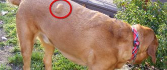
- Tumor on a dog's neck. A mass in the neck area may be a sign of the development of mastocytoma. This is a tumor consisting of transformed mast cells (immunoactive elements, tissue macrophages). Its danger lies in its rapid growth and negative impact on the immune system. Also, over time, compression of the esophagus, upper respiratory tract and main blood vessels is possible, which leads to death.
Tumor formation on the dog's neck
Types of bumps on the neck
Sometimes an owner, when examining a pet, discovers a purulent lump on the dog’s neck. If you are sure that the formation is benign in nature, you can help the animal yourself:
- It is necessary to carefully remove the pus from the wound. In this case, you should protect your own hands with disposable rubber gloves.
- Then rinse the area with a swab soaked in a disinfectant solution. Chlorhexidine, a pale pink solution of potassium permanganate, and chamomile infusion are suitable.
- Apply a bandage with wound healing ointment. Levomekol ointment is excellent for these purposes. It is sold in a regular pharmacy.
The wound needs to be treated once a day: rinse with a disinfectant solution and change the bandage. It is recommended to repeat these steps until complete healing.
Causes of a tumor on the face of an animal
There are many reasons why a tumor appears. It happens that they can be seen on one and the other half of the muzzle. This may develop due to:
- Infections/cellulitis;
- Allergies whose causes are difficult to determine;
- Poisoning with paracetamol or products containing it;
- Collections of liquid or clotted blood;
- Inflammation of muscle tissue;
- Inflammation of skeletal muscles;
Diagnosis
A detailed medical examination is carried out by a veterinarian to quickly establish the cause of the disease:
A tumor in a dog can be the result of an allergic reaction after an insect bite or taking paracetamol. The site of the tumor causes pain when palpated. As a rule, this is accompanied by elevated temperature and poor health of the animal. The formation grows to the tissue, but there are exceptions to the general rules.
The tumor develops as a hematoma after injury. In this case, only one side of the animal's face swells. The tumor, when pressed, can cause pain, and in some animals the area even turns blue.
Chondrogenic, osteogenic, lymphoid and fibroid sarcoma are types of malignant tumors in dogs that can appear on the face. Swelling of the lymph nodes located under the jaw can also appear as a tumor on the face. It can cause pain and appear as an open wound. Swelling is located in the area of the lower facial bone or in the upper area of the head.
Useful video
Tumors in female dogs: a tumor on the tail causes venereal sarcoma, uterine fibroids, fibromas, more often - the genitourinary vestibule, less often - the entrance part of the female genital organs. It is rational to carry out treatment only through surgery. In rare cases, after examination and a series of mandatory tests, the uterus and ovaries are removed.
Castration is one of the mandatory conditions of therapy, because all tumors that appear under the tail, on the genitals and mammary glands are associated with changes in hormonal levels. In addition, some tumor diseases that appear on or under the tail signal a sexually transmitted infection. Therefore, female dogs are prohibited from being bred.
Lipoma will make you wary
A painless formation on or under the tail, regardless of the sex of the animal, may turn out to be a lipoma or, as it is called, a wen. This, at first glance, is a harmless formation that grows extremely slowly and does not make itself felt; it will quickly develop into cancer.
But at the same time, the wen does not cause discomfort; it does not prevent the animal from running, sitting, or meeting its natural needs. In this case, it does not need to be treated. If a lipoma can become a liposarcoma, increase in size and interfere with the normal functioning of the animal, it is removed exclusively through surgery.
Lipoma often manifests itself in the form of a lump or papilloma that appears on the tail. To make a correct diagnosis and prescribe treatment, the doctor must carry out diagnostics. He examines the deep groin area and lymphatic system. The doctor takes an x-ray of the abdominal cavity and chest to check for the appearance of secondary growth lesions that appear in any malignant tumors in dogs. If during the research the cause of tumor formation could not be determined, the doctor proceeds with a biochemical analysis.
Benign neoplasms of the perianal (hepatoid) glands can be diagnosed by their types and the results of sampling the pathological formation for research and analysis. It is very difficult to diagnose the histological structural and functional formations of adenoma cells that develop from glandular cells.
But when making a diagnosis, the doctor takes the growth form as a basis, because the malignant type is distinguished by a tendency towards discreteness and local infiltration. Adenocarcinomas often form secondary foci of tumor development. A doctor will never make a diagnosis based on the appearance of a perianal adenoma; an excisional biopsy must be performed.
Any tumor on the tail should alert the owner and cause a visit to the veterinarian.
Tumors in the mammary glands in dogs
According to a number of statistics, this type of tumor makes up more than half of all cancers in dogs.
Mostly female dogs are affected by the disease, but only in one percent of cases is it diagnosed in males. Most often, breast formations appear at the age of ten, sometimes this can happen earlier. The disease is extremely rare in animals under four years of age.
The tumor is described as an uncharacteristic growth of iron tissue that cannot be controlled by the body. It contains atypical cells with an irregular structure. They differ from functional cells, both in their operating principle and in structure.
Cells with a different nature begin to quickly divide and transport their impulses throughout the genetic code. Tumor tissue grows rapidly by cell division. The process of how exactly a tumor arises and develops has been studied for a long time, but it is impossible to obtain more complete information about this process. This is due to the difficulties in conducting relevant research.
A malignant formation can develop up to several years without clinically manifesting itself. How quickly a specific tumor will grow and develop is personal in any case, so the prognosis for the animal should always be approximate from the very beginning.
Neoplasms on a dog's chest are often divided into benign and malignant. Among all tumors, more than half of them belong to the latter type. The most common is cancer.
A characteristic feature of malignant tumors is their metastasis; they can spread throughout the body through the bloodstream or lymph. Harmful cells that have spread throughout the body, in some cases, cause tumors to appear in other organs.
Cancer cells often cause the formation of secondary growth foci in the lymphatic system, and only then do they develop in the circulatory system. Therefore, the first signal of disease progression is the appearance of a secondary tumor in the closest lymph node.
For distant metastasis, the most likely area of spread of lesions will be the respiratory system, but growth in other systems is also likely. The fact that tumors are benign and also malignant is conditional, because any benign manifestation can, as a result of exposure to a carcinogen, develop into cancer.
Dental problems
If the bone formations are inflamed or broken, swelling may form on the face, causing fever, loss of energy, and discomfort for your dog. A course of dental therapy and antibiotics will help get rid of the formation. Your dog, like you, may begin to react to external factors that cause tissue inflammation. This:
- chemicals, changes in diet, flowering plants, insect or spider bites;
- medicines and a number of vaccines. A severe form of allergy can cause the formation of many tumors, which are localized not only on the face, but also in the larynx.
The basis of treatment should be the elimination of the factors that caused the allergic reaction and the use of pharmacological, antihistamines, steroids, and the use of antibiotic-based ointments. The dog must follow a diet until the end of the course of treatment.
Treatment of facial swelling in dogs
The proper treatment for facial swelling in dogs usually depends on the cause. It is important to seek your veterinarian's advice before beginning treatment.
Extreme allergic reactions and serious infections can cause the airways to swell, making it difficult to breathe. This is an emergency; Get to the nearest open veterinary center immediately.
Mild to moderate allergic reactions can be treated at home with medications.
The reaction is considered mild to moderate if your dog is breathing normally and acting relatively normally (no more than slightly lethargic). In these cases, your veterinarian may recommend giving an over-the-counter antihistamine (loratadine, Zyrtec). If your dog's face is swollen for any other reason (or for unknown reasons), the next step is to take your dog to the veterinarian for an examination. Your veterinarian will recommend treatment based on the diagnosis.
Swelling of the muzzle due to dental or oral problems may require professional dental work. The dog is often treated first with antibiotics and anti-inflammatory drugs and then placed under anesthesia and a complete dental cleaning and examination. Dental x-rays can be taken while your dog is under anesthesia. Depending on the cause, your veterinarian may need to remove teeth or perform another type of oral surgery. Your dog may be sent home with antibiotics to treat or prevent infection.
If the injury occurs on the face or head, treatment depends on the severity of the injury. X-rays may be needed to determine the severity. Start by administering any necessary first aid, then contact your veterinarian immediately. Treatment often includes antibiotics, anti-inflammatory drugs, and supportive care. Serious injuries may require surgery. Snake bites are treated with supportive care and sometimes antivenom (if available and necessary).
If your vet suspects a tumor and/or cancer, additional testing will be needed to find out more. Your veterinarian may recommend blood tests, x-rays, and analysis of the tumor itself (usually a fine-needle aspirate or biopsy sent to a pathologist for microscopic analysis). Treatment depends on the diagnosis. Cancer may need to be treated with chemotherapy, radiation and/or surgery.