Diseases of the vestibular system lead to some of the most dramatic manifestations observed in clinical neurology. The vestibular apparatus is responsible for the orientation of the animal depending on gravity. Consequently, vestibular dysfunction is reflected in abnormal posture of the body, including the head, limbs and eyes. The results are falls, incoordination, head tilt, nystagmus and ataxia. This article provides an overview of the most common diseases affecting the vestibular system in dogs and cats.
Anatomy
The anatomical structure of the vestibular apparatus is described in various textbooks (deLahunta, 1983). Simply put, the vestibular apparatus consists of receptor organs inside the ear that record the static position and movement of the head relative to the surface of the earth (gravity). To determine static position, vestibular receptors detect the movement of small crystals (statoconia) in the inner ear under the influence of gravity. Statoconia are located inside the gelatinous ottolith membrane. The cilia of the vestibular receptor cells protrude into this gelatinous membrane. The force acting on these statoconia causes the cilia to bend, which provides the central part of the vestibular apparatus with information about body position.
When the head moves, the movement of fluid (endolymph) in small tubular structures (semicircular canals) leads to the movement of cilia on additional receptor cells within the equilibrium ridges. This movement of the cilia excites receptor cells, which transmit this information through the vestibular nerve to the central parts of the vestibular apparatus. Thus, vestibular receptors collect information about head movements in space.
The nerve fibers emerging from these peripheral receptors form the relatively short vestibular nerve. Nerve fibers end in the vestibular nucleus or within the part of the cerebellum (cerebellar tent nucleus) associated with vestibular functions, so anatomical causes should be sought for cerebellar diseases associated with symptoms of imbalance.
Structure and functions of the vestibular apparatus in dogs
The vestibular apparatus is a complex of structures of the inner ear, united into a single system of semicircular canals, which respond to movements, turns, and tilts of the head. The channels are located at right angles and are filled with liquid.
The vestibular apparatus is located in the bone structures of the inner ear on both sides. Its basis is the accumulation of ciliated cellular structures of the inner ear, endolymph, and the work is carried out due to fluid pressure on the elastic membrane.
The main task of the complex receptor of the vestibular analyzer is to maintain the balance of vertebrates and the orientation of the body in space.
The vestibular apparatus is divided into two sections:
- Central. Located in the cerebellum, medulla oblongata.
- Peripheral. Located near the middle ear.
Clinical symptoms of diseases of the vestibular apparatus
Clinical symptoms of vestibular dysfunction are reflected in impaired orientation of the head, limbs and eyes. Head tilt, nystagmus, and ataxia are common, whether the disease affects the peripheral receptors (peripheral vestibular disease) or the central nuclei, cerebellum, or central pathways (central vestibular disease). The head is usually tilted in the direction of the disorder. However, if there is a disturbance in the inferior cerebellar peduncle, the head will tilt in the opposite direction. The localization of the disorder is often facilitated by ipsilateral hemiparesis, which will correspond to the side of the disorder.
Nystagmus (characteristic fast and slow eye movements) is often associated with vestibular dysfunction. Nystagmus may be normal (visual-vestibular response) when turning the head from side to side. The fast phase of eye movement will indicate the direction of head movement. This slow sliding and rapid return of the eye as the head moves from side to side is considered normal. In animals with bilateral lesions of the vestibular apparatus, the visual-vestibular reaction will be absent. When the vestibular system is dysfunctional, the eyes tend to spontaneously slide in the direction of the disorder (slow phase), and thanks to a homing mechanism in the brain stem, the eyes quickly return to their original position (fast phase).
Pathological nystagmus can appear spontaneously (in a resting position) or when the position of the head changes (positional nystagmus). In the latter case, nystagmus appears only when the veterinarian forcefully turns the head into an unusual position. The easiest way to obtain positional nystagmus is to place the animal on its back. The direction of nystagmus is determined relative to the horizontal axis passing through the eye section. With horizontal nystagmus, eye movements occur along this axis, with vertical nystagmus, along a perpendicular axis. With rotational nystagmus, the eyes move around an axis clockwise or counterclockwise.
The direction of nystagmus is determined by the direction of eye movement in the fast phase. This may make it difficult to detect the disorder because it may be in the direction of slow phase eye movement. In peripheral vestibular syndrome, the fast phase is directed in the opposite direction from the disorder. With central disorders, the direction of the slow phase relative to the side of the disorder may vary. The vestibular system is involved in limb movement, which is easily determined by stretching the ipsilateral limbs. In the absence of a vestibular response, ataxia or a fall may occur. The animal prefers to lie on the side where the disturbance is located. The ipsilateral limb usually has decreased extensor muscle tone, and the opposite limb, on the contrary, has increased extensor tone. The animal may walk in circles, usually in the direction of the disturbance. The central vestibular pathways typically include ascending and descending motor and sensory pathways to the extremities. Consequently, paresis is common. Because the influence of the vestibular system on the limbs will be ipsilateral, disturbances in the brain stem will affect the limbs on the same side as the disturbance. Normal vestibular control is also important for maintaining the eyes in a normal position within the socket. From the vestibular apparatus, information passes through the medial longitudinal fasciculus to the III, IV and VI cranial nerves. If the vestibular input is impaired, then when the head is turned to an aberrant position, a pathological position of the eyes (strabismus) may be observed. This is easiest to observe when the animal’s head is extended in a dorsal position. When viewed from above, ventral and ventrolateral strabismus can be seen on the eye on the affected side. The dorsal part of the sclera in this eye is more damaged than in the healthy eye. Vomiting and nausea are common in people with vestibular disease and are usually associated with peripheral vestibular disorders. Animals with vestibular dysfunction also vomit, especially when the vestibular system is acutely affected. Nausea is difficult to detect in animals, but it can manifest itself in anorexia, which is often observed in acute diseases of the vestibular system.
Symptom of Shifty Eyes in Dogs
- Enophthalmos is a displacement of the eye towards the orbit. The size of the eyeball remains the same; it seems to go deeper into the orbit;
- Microphthalmos – reduction in the size of the eyeball;
- Exophthalmos of the second eye - protrusion. In this case, it is possible to mistakenly assume that the healthy eye is “retracted”;
- Anophthalmos is a congenital absence of the eye, sometimes owners confuse it with microphthalmos;
We recommend reading: Why the Cat Tears
According to the nature of occurrence and duration of the flow, they are divided into:
- Congenital, chronic eye diseases;
- Acute, acquired;
- Chronic, acquired;
Causes of sunken eyes in dogs:
- Constitutional enophthalmos in dolichocephalic dogs. These breeds include greyhounds, the most common representatives of these breeds are: greyhound, Irish wolfhound, Italian greyhound, whippet, etc. It is a physiological norm - a feature of these dogs.
- Horner's syndrome is a neurological disorder that causes sunken eyes, constricted pupils, prolapse of the third eyelid, and drooping of the lower eyelid. More often it appears on one side. When the cause causing the development of Horner's syndrome is eliminated, the shape and position of the eyeball is completely normalized.
- Dehydration. With prolonged water deficiency or diseases accompanied by significant losses of fluid from the body, dehydration develops. And since the eyes are more than 90% fluid, a decrease in the total amount of fluid in the body by even 5% causes the dog’s eyes to sink. The lesion is bilateral, the condition requires immediate medical attention.
- Microphthalmos and anophthalmos are most often congenital pathologies. Microphthalmos is a small size of the eyeball. Anophthalmos is the absence of the eyeball. There is no treatment.
- Decrease in muscle volume (atrophy) and fat in the orbital area. It is observed during prolonged fasting, diseases associated with impaired muscle tone. Etc. It is necessary to identify the cause and prescribe treatment.
- Wrinkling (atrophy) of the eyeball most often develops against the background of chronic inflammation of the eye. The prognosis is guarded and treatment is aimed at reducing the rate of ocular atrophy.
- Trauma to the eyeball with perforation of the cornea and outward leakage of the vitreous leads to a decrease in the volume of the eyeball. Requires immediate medical attention. With timely treatment, it is possible to preserve the eye and vision.
- Adenoma of the third eyelid, prolapse of the lacrimal gland, neoplasms and abscesses of the eyelids (conjunctiva). These diseases cause the eyeball to press into the orbit. When the cause is eliminated, the eye retraction stops.
If you suspect that your dog has a sunken eye, you should definitely take it to the veterinary clinic in Krasnoyarsk to an ophthalmologist. since this symptom manifests itself in the presence of serious systemic diseases or eye diseases. To obtain good treatment results, it is very important to correctly and timely identify the cause of eye retraction. In our clinic, appointments are conducted by a narrow specialist - ophthalmologist Busel Marina Nikolaevna. Call at any time, our receptionists will try to find the most convenient appointment time for you.
Neuroanatomical location
To select diagnostic tests, it is necessary to differentiate vestibular syndromes into central - inside the brain stem and peripheral - VIII cranial nerve and its receptors (Table 1).
| Table 1. Differentiation of peripheral and central vestibular syndrome | ||
| Clinical symptom | Central | Peripheral |
| Nystagmus SpontaneousPositions | Horizontal Rotational Vertical Variable | Horizontal RotationalPermanent |
| Head tilt | Present | Present |
| Cranial nerve damage | In any, except VII | VII |
| Horner's syndrome | + /- | + /- |
| Proprioceptive disturbances of consciousness | Present | None |
Certain clinical symptoms are associated with central vestibular syndrome. Their absence, however, does not allow us to exclude this syndrome. Head tilt, horizontal and rotational nystagmus and ataxia can be observed in both central and peripheral vestibular syndrome. Positional vertical nystagmus and limb paresis most often indicate central vestibular syndrome. In unilateral central vestibular syndrome, hemiparesis is observed ipsilateral to the disorder. Sometimes hemiparesis is present on the side opposite to the direction of head tilt (paradoxical vestibular syndrome). In this situation, the disorder will be on the ipsilateral side of the hemiparesis.
In dogs with bilateral peripheral vestibular syndromes, head movement cannot evoke any visual-vestibular responses. These animals usually have a wide set of limbs. They hold their head low to the ground and can move it noticeably from side to side.
After localizing the disorder, the correct differential diagnosis should be formulated. Unfortunately, all intracranial disorders result in symptoms indistinguishable from peripheral vestibular syndrome. Conversely, animals with acute and severe vestibular dysfunction may be so weak that no neurological assessment can be performed. Because of these nuances, if the veterinarian is unsure of the location of the disorder, testing for peripheral and central vestibular syndrome should be conducted in parallel.
Practical advice
As mentioned above, treatment and preventive measures vary between diagnoses and also depend on the severity of symptoms and the dog's overall condition. Always follow your veterinarian's instructions exactly. However, in most cases, it is recommended to re-check the pet’s condition two weeks after the initial treatment and start of treatment in order to track the positive dynamics and, if necessary, adjust the treatment. Secondary symptoms, such as dehydration due to vomiting , should also be monitored and addressed. Complete recovery is not possible in all cases, it all depends on the severity of the disease and its causes.
Peripheral vestibular syndrome
Idiopathic peripheral vestibular syndrome is observed in dogs and cats (Schunk, 1990). Most often, old dogs (geriatric vestibular syndrome) and young and middle-aged cats are affected. In the northeastern regions, cats often get sick at the end of summer. No reasons were identified. The cause of the disease in cats in the southeastern regions is believed to be that cats eat the tails of blue-tailed lizards. Clinical symptoms of acute peripheral vestibular syndrome are nystagmus (horizontal and rotational), head tilt (in the direction of the disorder), rocking gait and falls. No other neurological symptoms are observed. Initially, clinical symptoms are very severe. For Horner's syndrome and facial nerve paresis, other differential diagnosis points should be considered. Differential diagnoses for peripheral vestibular syndrome include otitis media of the inner ear (dogs and cats), middle ear polyps (cats), and neoplasia (squamous cell carcinoma in middle-aged cats). It is necessary to carry out otoscopy, x-ray of the tympanic bladder and other modern studies - computed tomography (CT) and magnetic resonance (MR).
Clinical symptoms of idiopathic vestibular syndrome disappear quickly within 1-2 weeks. Nystagmus disappears first (during the first days). Improvements in body posture and gait are observed over 5-7 days, but slight head tilt may persist. Although most animals achieve full compensation, some may experience temporary ataxia, especially after jumping upwards. There is no cure, and relapses are possible.
Otitis media/interna is a common cause of vestibular dysfunction. It most often develops as a result of a bacterial infection that spreads from both the external auditory canal and the pharynx through the auditory tube. Less commonly, the infection spreads hematogenously. Foreign bodies such as grass awns can predispose to severe infection.
Rice. 1. Contrast T-magnetic resonance scan of the skull of a dog with external otitis in a cross section. A contrast-enhanced lesion was noted in the right tympanic bulla (small arrows). The external auditory canal is also highlighted (large arrow). Histological diagnosis is chronic infection due to migration of a foreign body (grass awn)
Clinical symptoms may reflect primary vestibular or auditory dysfunction and involvement of the external ear. Often there is soreness in the area of the outer auricle and pain when opening the mouth. It is expected that more than half of animals with otitis media/interna will also have facial nerve involvement. Otoscopy is used to examine the eardrum. This is a difficult procedure for animals with severe otitis externa. With middle ear disease, the eardrum changes color, becomes hyperemic, opaque, and bulges outward. A clear or yellow liquid is visible behind the membrane. X-rays of the tympanic bladder and other modern imaging methods are also used to confirm the diagnosis (Fig. 1). An accurate diagnosis is made by the results of bacterial cultures taken through myringotomy or during surgical exploration.
Rice. 2. Otoscopic examination of the dog’s external auditory canal with the head tilted. A clear liquid can be seen covering the mass. Histological diagnosis - inflammatory polyp
Ear tumors are most common in older animals. The most common are squamous cell carcinoma and adenocarcinoma. Inflammatory polyps occur in cats. During otoscopy, tumors can be seen extending beyond the eardrum (Fig. 2). Cranial X-rays and other techniques are required to visualize the middle and inner ear. However, the abnormalities observed in these images do not always correspond to neoplasia, so studies of tissue taken during surgical examination are necessary to clarify the diagnosis. Destruction (lysis) of the bulla bone is most often associated with neoplasia rather than inflammation.
Treatment is surgical resection/reduction of the tumor body, radiation and chemotherapy.
Congenital peripheral vestibular syndrome occurs in German Shepherds, Dobermanns, English Cocker Spaniels, Siamese and Burmese cats. While peripheral vestibular syndrome is most often an idiopathic condition, congenital peripheral vestibular syndrome in young Dobermanns is associated with lymphocytic labyrinthitis (Forbes and Cook, 1991). Bilateral congenital peripheral vestibular syndrome occurs in Beagles and Akitas. Clinical symptoms include ataxia, head tilt, and sometimes deafness. Symptoms may persist throughout life or resolve spontaneously. No treatment has been developed. Metronidazole poisoning can result in symptoms of central vestibular syndrome in dogs and cats (Dowetal 1989; Saxon and Magne 1993). This usually occurs when high doses of this drug are given. Since metronidazole is eliminated through the liver, toxic levels of the drug may be present in the serum of animals in certain liver dysfunctions. The first clinical symptom will be ataxia, progressing to nystagmus and more severe vestibular disturbance. Clinical signs most often reflect central vestibular dysfunction, and abnormalities are found in the brainstem in dogs. If the serum concentration of metronidazole is measured immediately after the onset of clinical symptoms, it will be at a toxic level. If there is a delay in determining the serum concentration of metronidazole, it will soon return to normal, but clinical symptoms will remain.
There is no specific treatment for metronidazole poisoning. The main measure is to stop using the drug. With severe initial clinical symptoms, some dogs may die. Others recover completely in 1-2 weeks.
Aminoglycosides, given systemically or topically, can cause vestibular symptoms and deafness. Streptomycin and gentamicin do not have a pronounced effect on vestibular receptors, but neomycin, kanamycin and amikacin damage mainly auditory receptors. Chlorhexidium solution, which is used to cleanse the external auditory canal, can also lead to vestibular disorders. In addition, other idiopathic and inflammatory neuropathies can affect the vestibular nerve. These diseases are not well understood, so it is very difficult to make a differential diagnosis. A similar situation exists in the case of certain metabolic disorders such as hypothyroidism and vestibular neuropathy. It is not always possible to establish the cause and effect of these diseases.
Provoking factors
Among the reasons that provoke the development of vestibular syndrome in dogs are:
- previous severe traumatic brain injury;
- inflammation of the middle or inner ear;
- disorders of hormone synthesis (thiamine deficiency);
- uncontrolled use of antibiotics based on aminoglycosides (amikacin, neomycin, geomycin, which some dog breeders use for self-medication due to their low price);
- mengoencephalitis;
- neoplasms of the inner ear (cysts, polyps, tumors);
- excessive use of ear cleaning products;
- autoimmune processes due to which the body “attacks” its own nerve tissue.
On a note! The disease can develop in dogs of any gender, age and breed. But most often the diagnosis is made in Dobermans, cocker spaniels, beagles, German shepherds, fox terriers and Tibetan terriers.
Central vestibular syndrome
Tumors in the infratentorial space, such as meningioma, and choroid plexus tumors may cause vestibular symptoms due to infiltration or compression of the vestibular nerve (Figure 3). Meningiomas can form flat-shaped lesions. Tumors of the choroid plexus grow around the fourth ventricle, often at the level of the lateral apertures. Diagnosis of intracranial neoplasms is carried out using modern imaging methods. Abnormalities and associated brain structures are better seen with MR than with CT, since in the latter case radiation artifacts often obscure structural details in this field. Surgical reduction or resection of the tumor is the ideal treatment, but this is often hampered by the insensitivity of the tumors and the proximity of vital brain structures. Radiation can be used to slow tumor growth, but choroid plexus tumors are relatively resistant to radiation.
Rice. 3. Preoperative contrast T-magnetic resonance scan (A) of a dog with the head tilted to the right. A contrast-enhanced formation is visible located in the right corner between the cerebellum and the pons (arrow). B. Postoperative contrast-enhanced CT scan (CT) at approximately the same level shows resection of the mass (arrow). Histological diagnosis: meningioma
The most common nutrient deficiency that affects the central nervous system is thiamine deficiency. It is more often observed in cats and leads to damage to the nuclei of the optic and vestibular nerves, the caudal eminence and the lateral geniculate ganglion. The first clinical symptoms are vestibular ataxia, progressing to seizures with ventral flexion of the neck and dilation of the pupil and complete absence of response to light. Treatment for such deficiency is the administration of thiamine, parenterally and intravenously.
Inflammatory diseases can also affect the brain stem and other parts of the nervous system. They can have an infectious and non-infectious etiology. The incidence of infections associated with meningitis varies depending on geographic location. For the majority (60%) of meningitis syndromes in pets, an infectious cause cannot be clearly identified. Infectious agents. causing brain diseases can be viruses (distemper, parvovirus, parainfluenza, herpes, feline infectious peritonitis, pseudorabies, rabies), bacteria and rickettsia (Rocky Mountain spotted fever and Ehrlichia), spirochetes (Lyme disease, lentospirosis), fungi (blastomycosis, histoplasmosis, crintococcosis, coccidioidomycosis, asnergillosis), protozoa (toxonplasmosis, neosnorosis) and unclassified organisms (nrotothecosis).
Specific rickettsiae that cause Rocky Mountain spotted fever. affect the brain stem, and especially the vestibular apparatus (Greeneetal., 1985).
There is usually a history of systemic disease (accompanied by thrombocytopenia) that occurred 5-10 days before the development of neurological symptoms. As the fever decreases, neurological symptoms appear. The scan does not detect intracranial formations. Sometimes in sick dogs, contrast enhancement is noted in the area of the choroid plexus. It must be differentiated from normal contrast enhancement in this structure. Cerebrospinal fluid usually contains a slightly increased number of nucleated cells (less than 50 nucleated cells/µl, normal - less than 5 nucleated cells/µl) and a slightly increased protein concentration (less than 50 mg/dl; normal - less than 25 mg/dl). The diagnosis is confirmed by an increase in the titer of antibodies to the microorganism, but these results are often obtained after the disease has progressed. The prognosis depends on the severity of clinical symptoms before treatment. Dogs with severe depression are unlikely to recover. Therefore, dogs with vestibular syndrome following a systemic illness accompanied by fever and thrombocytopenia should be treated with tetracycline and doxycycline, followed by a definitive diagnosis based on antibody titers.
Animals often suffer brain injuries from being hit by a car. Brainstem function is assessed by the functions of the cranial nerves, especially the visual-vestibular reflexes. Sometimes dogs with cranial and cervical disorders develop symptoms of brainstem disorders, therefore, all manipulations to identify reflexes should be carried out only after assessing the stability of cervical fractures and dislocations. Otoscopy may reveal bleeding in the ear canal.
The diagnosis is confirmed by obvious trauma. X-rays show skull fractures. Modern imaging techniques are used to assess intracranial bleeding and edema. In the first 12 hours after severe trauma, it is best to use CT to detect bleeding. Treatment is based on the identified pathophysiological consequence of traumatic brain injury - cerebral edema. To stabilize intracranial pressure, surgical debridement of bleeding is often required. Vascular diseases causing central vestibular syndrome and associated with the cerebellum are rare. The use of modern imaging methods makes it possible to make an intravital diagnosis.
Methods of diagnosis and treatment
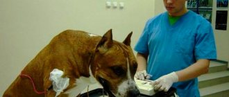
At the first symptoms of your pet, it is important to take it to a qualified veterinarian. For a correct diagnosis, you will need a card with the dog’s medical history and a number of diagnostic tests:
- urine and blood tests;
- X-ray diagnostics to assess the condition of the middle and inner ear;
- MRI, CT to examine the presence of structural changes in the brain;
- tests to determine the reaction of the nervous system to stimuli;
- ear scraping;
- Ultrasound diagnostics of internal organs;
Treatment of vestibular syndrome is selected taking into account the causes that provoke the pathology. For the idiopathic form (congenital or senile), specific treatment has not yet been developed. The veterinarian can only select medications that alleviate the animal’s condition, and the symptoms of the disease often decrease 72 hours after the onset; within a week the dog can move independently, but the likelihood of reoccurrence of the pathology remains.
Depending on other causes, the following treatment options may be used:
- For inflammatory processes associated with infections, broad-spectrum antibiotics are used.
Important! In case of otitis, it is strictly forbidden to use ototoxic drugs with aminoglycosides (antibiotics, chlorhexidine) for a dog.
- If oncological pathologies are detected, surgery or a course of chemotherapy is prescribed.
- For endocrine disorders (hypothyroidism, etc.), replacement therapy is used.
- Physiotherapeutic procedures are used as an additional support.
A timely diagnosis and proper treatment make it possible to stabilize the dog’s condition already on the 2-3rd day, and after 14 days to get rid of the head tilt to a much greater extent. To avoid relapses, treatment should not be stopped at the first visible improvement in the condition. The extent and time of recovery can vary from several weeks to several months, and some animals remain with a slight head tilt for life.
Diagnostic tests
If the disorder is suspected to involve central vestibular structures (forebrain, brainstem, or cerebellum), then modern imaging techniques (CT and MR) are used to assess the structural integrity of the brain. This examination is non-invasive but requires anesthesia for all animals except comatose ones. Conventional x-rays are used for skull fractures and diseases of the middle ear (tympanic bladder), but they do not evaluate the parenchyma of the nervous system. If peripheral vestibular syndrome is suspected, the main method of evaluation is otoscopy of the animal under anesthesia. Analysis of cerebrospinal fluid (CSF) is necessary mainly for the diagnosis of inflammatory diseases. The most accurate diagnostic method is to collect CSF caudally to the site of the disorder. The liquid is analyzed for cellular composition, protein content and cell morphology. Although CSF analysis often aids in the diagnosis of diseases of the nervous system, it does not by itself lead to the establishment of an exact etiology. Antibody titers to specific infectious agents are measured in the CSF to assess intrathecal antibody production. However, if the blood-brain barrier is disrupted, antibodies can pass into the CSF from the central circulatory system. In such a situation, correlation of CSF and serum titers is necessary. Elevated antibody titers in CSF compared with serum suggest local antibody formation in the central nervous system and, therefore, overt CNS infection. CSF protein electrophoresis can provide additional information about the integrity of the blood-brain barrier and local immunoglobulin production.
Auditory brainstem potential (ABP) recordings are necessary to determine the integrity of the auditory pathways and can also provide information about the central pathways (brainstem) associated with hearing (Steisse et al., 1994; Fisher and Obermaier, 1994).
For intravital diagnosis of intracranial disease, surgical biopsy is often required. It is very difficult to perform in the infratentorial space because surgical exposure will be incomplete, especially for ventrally located lesions. Surgical exposure of lesions located at the angle between the cerebellum and the pons may increase occlusion of the overlying transverse sinuses. Limited access to these areas often prevents complete resection of the disorder.
For lesions of the auditory canal and tympanic bulla, lateral resection of the auditory canal and osteotomy of the tympanic bulla are used, respectively, for biopsy, resection of the disorder and drainage of infected tissue. If this procedure is performed to evaluate animals without vestibular symptoms, head tilt, ataxia, and nystagmus may result from damage to the vestibular structures.
Nystagmus in a cat - why does a cat's eyes run around?
Just like humans, cats' eyes are a reflection of their health and state of mind. When your cat's eyes shift unusually, this is a sure sign that there is a problem with the nervous system. Rhythmic vibrations of the eyeballs (nystagmus) occurs in both cats and dogs. The animal's eyes twitch involuntarily, the pet is unable to control the vibrations.
We recommend reading: Siberian Marble Cat Breed
Types of nystagmus in cats
There are two types of nystagmus - jerking and pendulum. Jerk (jerk) is characterized by slow eye movements in one direction, and then a sharp return to the previous state. With pendulum nystagmus, slight fluctuations of the pupils occur, in which the eyeball almost does not move. However, the jerk type is much more common in veterinary practice.
There is another classification of nystagmus - horizontal and vertical (according to the plane of eye oscillation). The vertical type occurs only with deep brain lesions and is a minor symptom. In other disorders of the nervous system, the most common is the horizontal type.
There is one more important circumstance. So-called congenital nystagmus often occurs in Siamese cats. In most cases, this is not a pathology, but a normal physiological reaction that allows you to adapt to the environment. The same can be observed in people who have overloaded their vestibular apparatus (see below).
Symptoms of nystagmus
In addition to the main symptom of “shifty” eyes, your pet may also turn its head and even spin in place.
Causes of nystagmus in cats
Most of the reasons for “shifty” eyes lie in the nervous system. Disorders of both the peripheral and central nervous systems can lead to the development of nystagmus. Shifty eyes are often associated with problems with the vestibular system, which is sensitive to the balance of the head and body.
Examples of causes of peripheral nerve diseases that lead to nystagmus include hypothyroidism, neoplastic tumors, and physical damage resulting from trauma (car accident, etc.). Diseases of the central nervous system can occur due to: thiamine deficiency (vitamin B1), tumor, viral infection (particularly feline infectious peritonitis), inflammation, heart attack, heart hemorrhage, toxin poisoning (lead and others).
Severe stress can also cause short-term nystagmus. This condition includes motion sickness in transport, as well as childbirth. Over time, the cat's vestibular system stabilizes.
Diagnosis of "shifty" eyes
Disturbances in the functioning of the nervous system can most often be diagnosed using CT (computed tomography). Sometimes a urine test is done to check for infections. If these procedures do not lead to any result, they resort to the most complex method in very prestigious clinics - analysis of cerebrospinal fluid.
Treatment of nystagmus
Treatment directly depends on the disease that caused the nystagmus and its severity. Only by getting rid of the main problem can this symptom be eliminated along with it. Diseases of the central nervous system will have to be treated more intensively than lesions of the peripheral nervous system. If your cat has anorexia and vomiting, you will need to give a special fluid by mouth to prevent dehydration. However, the list of medications for each case is individual, and the veterinarian draws it up based on the diagnosis. The same goes for aftercare.
Most neurologists recommend examinations every two weeks to monitor the progress of treatment. Secondary symptoms such as vomiting and dehydration should also not be ignored. Experience shows that cats recover faster after diseases of the peripheral nervous system.
Prevention of "shifty" eyes
It is impossible to formulate a specific system of preventive measures for nystagmus, because There are many reasons, including congenitality. The only thing we can advise is to avoid poisoning your cat with lead and other toxins. In addition to Siamese cats, cats with albinism are predisposed to this disease, keep this in mind. Some kittens under one year of age may exhibit nystagmus, but this is very rare and will go away soon.
There is another classification of nystagmus - horizontal and vertical (according to the plane of eye oscillation). The vertical type occurs only with deep brain lesions and is a minor symptom. In other disorders of the nervous system, the most common is the horizontal type.
Treatment
Specific treatment can only be recommended after an accurate diagnosis has been made. If intracranial tumors are diagnosed, then specific treatment is used - tumor reduction/resection and radiation therapy. In primary inflammatory diseases, if possible, it is necessary to identify the etiological microorganism, then specific treatment is used aimed at destroying the pathogen. In Rocky Mountain spotted fever, vestibular symptoms are treated with tetracycline and doxycycline. For toxoplasmosis, the combination of clindamycin and trimethoprim/sulfadiazine improves the condition and even eliminates clinical symptoms. Non-infectious inflammatory diseases of the central nervous system initially respond well to treatment with corticosteroids. Radiation therapy is used to treat granulomatous meningoencephalitis.
Nonspecific treatments include protecting the eyes from injury, especially when there is a facial nerve deficiency or when the animal is rolling on the ground or lying on its side. To reduce anxiety and anorexia, and sometimes to reduce head tilt and nystagmus, antihistamines such as diphenhydramine (1-2 mg/kg po or IV every 12-24 hours) are used.
Treatment regimen
The treatment regimen depends largely on the form, stage of the disease, the age of the dog, and the root cause. Prescribed individually by a veterinarian based on diagnostic results.
Treatment of VS in veterinary medicine includes:
- Drug therapy;
- Physiotherapy.
- Replacement therapy (for hypothyroidism);
- Rehydration.
Dogs are prescribed intramuscularly, intravenously, in tablets, glucocorticoids, antiemetics (dimenhydrinate), sedatives (diazepam), broad-spectrum antibiotics (clavulonate, amoxicillin) when diagnosing otitis media and viral infections.
In severe cases, if the dog cannot move independently, does not orient itself in space, loses its balance, hospitalization is carried out, and maintenance therapy is prescribed aimed at improving the condition and stopping the acute stage of the pathology.
Maintenance therapy is prescribed for the congenital form of the pathology, for old, elderly dogs. Surgical methods are used extremely rarely, for example, in the case of oncology.
Symptoms
As we noted above, the symptoms of vestibular syndrome in a dog can be similar or even identical to other serious diseases. For this reason, it is always necessary to be on alert. So what can indicate the development of this particular disease in a beloved “family member”?
- The defining symptom is a violation of spatial orientation. At the slightest sign of it, be sure to seek advice from specialists. As with any illness, in the early stages it is much easier to cope with the problem. By the way, a constantly or periodically tilted head, rotation around an axis, or stumbling is not a dog’s desire to play with others, but a sign of impending trouble.
- If you see twitching of the eyes, as in a nervous tic, the so-called vertical nystagmus, then this can also be (and in the vast majority of cases it is) a peripheral syndrome in dogs.
- Excessive salivation can be a sign of many diseases or even just some discomfort in your pet. But coupled with other symptoms, this should always be alarming.
- The same can be said, perhaps, about profuse gag reflexes.
Treatment, depending on the causes, degree of development and condition of the patient, is prescribed exclusively by a doctor. If a peripheral syndrome develops against the background of other diseases and is a side effect, then the root cause is initially or simultaneously eliminated.
In many cases (age-related and congenital), treatment is impossible today; drug therapy is used to alleviate the disease. And most importantly, do not self-medicate, as this will most likely lead to death.
Content
1. A little about the diversity of vestibular syndrome in dogs 2. About the catalysts for the development of the disease 3. Symptoms 4. Prevention
It's always a pleasure to watch your beloved dog scamper across the lawn, happily wagging his tail and jumping over obstacles. A healthy pet is a happy owner. But it often happens that coordination of movements is seriously impaired. And the reason for this is vestibular syndrome in dogs. When it occurs, there is an imbalance in the body, gait, and neurological deviations from the norm appear.
It is worth immediately noting that in the vast majority of cases, problems of this kind manifest themselves in elderly animals, as well as in people. And in this case it is useless to treat the disease - you can only neutralize its manifestation to some extent, achieving serious recessions. In puppies and young dogs, this pathology manifests itself less frequently, but it is also faster and easier to treat.
Signs and symptoms
Often, manifestations of the syndrome can be mistaken for a stroke. The main sign of pathology is loss of coordination in space. It is difficult for the dog to move, it walks with its head bowed, stumbles, and spins around itself. Some owners perceive this behavior as an attempt to play, and are in no hurry to take the dog to the veterinarian. This could cost her not only her health, but also her life.
Other symptoms of vestibular syndrome:
- vertical nystagmus (eye twitching),
- profuse drooling,
- uncontrollable vomiting,
- dizziness,
- bad response to voice
- paralysis of the facial muscles is possible.
In severe cases, the animal refuses to eat and empties itself.
Treatment methods
If a dog has congenital vestibular syndrome or it developed in an elderly dog (idiopathic form), then treatment in such cases is ineffective. The doctor can only prescribe medications to alleviate the condition and relieve some syndromes. If the pet becomes helpless, the owner will have to constantly look after it, turn it over, and massage it to avoid bedsores.
The syndrome, which developed against the background of inflammatory and infectious processes, requires the use of broad-spectrum antibiotics and anti-inflammatory drugs. In severe cases, corticosteroids (Prednisolone) are prescribed. If otitis is detected, it is prohibited to give the animal ototoxic drugs from the group of aminoglycosides, Chlorhexidine.
On a note! If the pathological condition arose as a result of a malignant formation, it can only be eliminated surgically. For hormonal disorders, replacement therapy is usually carried out.