If you notice how your pet constantly scratches the area in the eye area, and also has swelling of the eyelids, tearfulness is the first signal of the presence of a pathology of the organs of vision. Carefully examine the animal, or better yet, with such indicators of deviation from the norm, take it to a veterinary clinic.
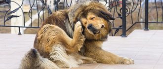
Dog scratching its eyes
Why does my dog constantly scratch his eyes?
The inflammatory process is always associated with itching and discomfort that bother the dog. The pet begins to behave irritably, actively scratching the area of the organs of vision, damaging the skin and the eye itself. The inflammatory process of the organs of vision is most often caused by mechanical damage during the walking period: the ingress of a piece of grass, a splinter, small fragments of glass, can also provoke a blow from a branch or while playing with other animals. Very often, the inflammatory process is caused by conjunctivitis or an infectious disease, for example, an inflammatory process of the liver, viral etiology (various groups of hepatitis), a highly contagious bacterial infection - plague, an infectious disease caused by mycoplasma bacteria - mycoplasmosis. When a dog’s body is infected with certain parasites, it also happens that the eyes become inflamed. Such diseases manifest themselves as general lethargy, loss of appetite, high fever, and apathetic behavior. Inflammation occurs simultaneously in both eyes.
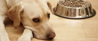
The clarity and purity of a pet’s gaze is an indicator of the absence of pathological changes in the visual organs
Causes of squinting
Common reasons include:
- Pink eye is a fairly common eye infection in dogs of all breeds. With this disease, squinting is a mandatory factor in the recovery of the eye, as it reduces pain and promotes eye hydration;
- Injuries to the organs of vision. Not a single animal is insured against such a circumstance, so injuries can occur both during a walk, for example, mechanical damage from a blow from a branch or an abrasion received in a fight with other animals, or the appearance of defects at home from bruises; Entry of a foreign object;
- Diseases of other kinds, for example, dry eye syndrome, corneal ulcer, blepharitis, keratitis, uveitis and the like;
- Allergic reactions to certain substances used in the home (deodorant, detergent or air freshener);
- Lens luxation;
- Inflammation of the tissues around the eyes;
- Exophthalmos;
- inversion of eyelids;
- Districhiasis;
- Thermal and chemical burns of the organs of vision.
Puppies or young dogs often have congenital pathologies, mechanical or infectious eye damage, and in adults and old animals - inflammatory diseases or those associated with weakened immunity, the appearance of neoplasms and tumors. If you notice a dog with a squinted eye, it needs mandatory supervision, since the animal’s behavior can clearly indicate a particular disease.
What measures to take when squinting is detected?
In cases where a dog squints its eye, the owner needs to do the following: Take the animal to a veterinary clinic and show it to a specialist, since only a doctor can correctly diagnose the pathology and formulate a therapy based on the examination and tests. The examination requires special education and equipment, because the scope of eye diseases is as broad as possible.
Carefully monitor your pet's behavior. A passive lifestyle, lethargy, refusal to eat, refusal to walk and play - all these are secondary symptoms that should alert owners. Moreover, squinting may only be a side effect of more serious pathologies accompanied by sensitivity to light.
Avoid walking in areas with direct sunlight. The best time for walking is in the afternoon or when the sun goes down. Walking in cloudy weather is also good, but the main thing is not to allow it to come into contact with your pet’s eyes when precipitation begins.
What does diagnosis suggest?
A diagnosis and a course of treatment can only be made after a complete veterinary examination of the dog (history and physical examination). After which the examination should be carried out by an ophthalmologist. He will check the eyelids, posterior and anterior chambers of the eyes, fundus, cornea, and also conduct tonometry and various tests of the visual organs, which will determine the level of tear fluid production. For some pets, examination is possible only after the administration of anesthetics or sedatives, which will protect the doctor and owner from defensive aggression on their part. In some cases, animals should undergo ultrasound and cellular analysis, as well as computed tomography or magnetic resonance imaging of the head. If more serious indicators are detected, you should donate blood and measure your blood pressure.
Causes of eye inflammation in dogs
The reasons why inflammatory processes in the organs of vision develop:
- An infection of an infectious nature is the spread of pathogenic microorganisms, both in the body itself and at the site of infection of the eye itself.
- A non-infectious lesion is a violation of the mechanical integrity of the organs of vision, neoplasms, ectropion of the eyelid, trichiasis of the ciliary margin.
- Genetic pathologies are deviations of intrauterine development, and may also be associated with the characteristics of the breed, as a result of selection.
If eye inflammation is expressed as a symptom of an ophthalmic disease, then after visiting a veterinarian, it can be cured by following the instructions and prescription:
- blepharitis;
- conjunctivitis;
- third eyelid prolapse;
- eyelid dermatitis;
- injury;
- neoplasm.
Only a veterinarian can determine the exact cause and prescribe treatment.
With these types of disease, only the eyes are affected. Otherwise, the animal behaves actively and nothing else bothers it. Drug treatment prescribed by a doctor quickly eliminates the disease and the dog is healthy again.
If it is accompanied by other symptoms, and the inflammation does not subside within three days, then this may be due to the spread of the virus. Therefore, it is necessary to regularly vaccinate your pet to prevent the occurrence of the virus in the blood.
The presence of parasites in a dog’s body can also lead to eye inflammation. Therefore, we should not forget that helminthiasis affects not only the gastrointestinal tract. Such parasites can easily enter the heart, lungs and other organs, and the eyes are no exception. Unfortunately, it will not be possible to carry out treatment on your own, so the dog will need to be shown to a veterinarian to assess the situation, and if the eye tissue is damaged, surgical removal of the dead helminths will be performed.
Video - First signs of eye disease in dogs
Symptoms of major diseases
Symptoms of eye disease are largely related to the age group, general health, body stability, frequency of negative influences, cause and stage of infection. Symptoms may include:
- discharge of pus, lacrimation;
- accumulation of fluid in the corners of the organs of vision (exudate);
- formation of scabs and scabs on the skin of the eyelid;
- swelling and redness of the eyelid;
- itching and burning;
- increased blinking frequency;
- scleritis, leukoma, opacity of the corneal layer;
- increased body temperature, weakened and lethargic state of the animal;
- impaired pupillary reactions in light, photophobia;
- blepharospasm;
- decreased vision.
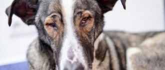
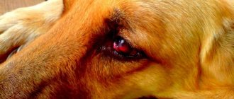
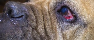
Pus discharge from a dog's eye indicates a disease
What you need to know! Be sure to take your pet to the veterinarian if you notice these symptoms. After diagnosis and examination, the specialist will make a diagnosis and prescribe the necessary medications.
Types of visual pathologies in dogs
Blepharospasm
Involuntary contraction of the orbicularis oculi muscle (blepharospasm) - repeated contraction of the muscles of the eyelid. The dog begins to avoid light, and the eyes secrete exudate. The eyelid swells and becomes painful.
Keratitis
Keratitis is an infection and inflammation of the cornea due to damage. Keratitis is often caused by infectious pathogens such as bacteria, adenoviruses, fungi, and thelaziosis (helminths). The dog begins to squint his eyes, scratch his eyes with his paws, become nervous and behave restlessly.
Keratitis is very dangerous and can lead to serious complications if not treated correctly.
Prolapse
Prolapse is a deviation in the placement of the lacrimal gland of the third eyelid. After the loss, inflammation and swelling occur. A characteristic feature of this deviation is that it can appear and go away after some time.
It is believed that prolapse has a hereditary predisposition
Conjunctivitis
Conjunctivitis is an inflammatory process of the film membrane (conjunctiva). The animal blinks frequently, constantly touches its eyes with its paws, and whines. In most cases, it has a viral and bacterial form.
Conjunctivitis is the most common eye disease in dogs.
Cataract
Cataract is a pathology of the light-refracting structure of the eye, characterized by clouding of the lens and loss of its transparency. During clouding of the lens, the dog is careful in movement and completely switches to auditory and olfactory perception. The main reasons for its occurrence can be considered heredity and old age.
Cataracts in dogs develop in old age and are often inherited
Blepharitis
Blepharitis is an inflammation of the skin tissue of the eye due to infection, insect bites, the presence of parasites, and burns of various types.
Inflammation of the eyelids occurs against the background of allergic reactions, injuries, infections
Entropion and eversion of the eyelid
Eversion of the lower eyelid (ectropion), inversion of the eyelid (entropion) - inversion and inversion of the eyelid, a deviation that mainly predominates in dogs. Soreness appears, the conjunctiva dries out, purulent discharge and excess tear fluid occur.
Pathological changes in entropion and ectropion of the eyelid occur in the eyelid itself in breeds with folded skin and a large palpebral fissure
Dermatitis of the century
Dermatitis of the eyelid – provokes the occurrence of pathologies. The tissues acquire a reddish tint, peel, pus is released, swelling appears - all this actively develops conjunctivitis and keratitis.
Dermatitis affects the tissue around the eye and provokes the active development of other diseases
Ulcerative keratitis
Ulcerative keratitis. After the inflammatory process, the affected corneal tissue becomes thinner, and small ulcers occur. The contours of the pupil blur, the cornea gradually becomes cloudy, and the membrane of the eyeball acquires a red tint. The dog actively scratches its eyes, lacrimation and pus appear.
Damage to corneal tissue by ulcers
Lens luxation
Lens luxation is a dislocation of the lens from its normal position. Injury to the pupil and movement to another place, which often leads to deformation of the eyeball itself. It occurs after a complication of an infectious disease, injury, glaucoma, cataract and is often hereditary. Basically, complete loss of vision occurs.
Displacement of the pupil from the center to the side
Eyeball prolapse
Eyeball prolapse is a dislocation of the organ of vision beyond the eyelid. Appears due to cranial damage, intracranial pressure or muscle tension. More often, infection occurs with subsequent loss of vision.
Strong extension of the eyeball beyond the eyelid
Eye diseases
Other eye diseases in dogs: main symptoms and treatment methods
Eye diseases in dogs can be of 3 types:
- Infectious – occurs when infected with viruses or bacteria as a primary disease or complication of infection of another organ.
- Non-infectious - mechanical damage, neoplasms, inflammation due to improper growth of eyelashes, eversion of the eyelids.
- Congenital - characteristic of some breeds, for example, Shar Peis. Includes eversion, entropion of the eyelids, deformations of the eyes and lenses. They are usually corrected surgically.
Blepharospasm
Blepharospasm is a voluntary rapid contraction of the muscle tissue of the eyelids. It is quite simple to recognize this disease in a dog by rapid blinking, almost without stopping. In addition, the area around the eyes and the eyelids themselves swell noticeably. Touching them causes pain in your pet.
Exudate—liquid—accumulates in the corners of the eyes. Photophobia is observed - the animal begins to hide from light sources, go into the shadows on the street, constantly lowers its head and squints.
The pathology is not dangerous in itself, but indicates the presence of infection or inflammation in the body. Often the cause is spasm of the trigeminal nerve. Treatment is selected based on the original source. You cannot choose therapy for blepharospasm on your own.
Conjunctivitis
Conjunctivitis is the most common eye infection in dogs. It is expressed in dysfunction of the conjunctival mucosa and is often infectious in nature. In addition, the causes may be allergies, viruses, clogged tear ducts, foreign body trauma, irritation due to drooping of the eyelids or ingrown eyelashes.
The symptoms are pronounced: the conjunctiva turns red, swells, discharge of tears and pus appears, the third eyelid may swell, mild squinting and frequent blinking may occur. The dog begins to rub his eye with his paw, becomes restless and whines.
Treatment includes antibacterial drugs, application of tetracycline ointment and instillation of special drops.
Ectropion and entropion
Ectropion and entropion are the medical names for eversion and entropion of the eyelids. Both diseases usually develop in parallel, so they are practically not separated. They are expressed in a change in the shape of the eyelids (turning outwards or inwards), loss of their natural functions. When eversion occurs, the eyeball is left without moisture and protection from germs. When curled, the eyelashes irritate the pupil and can grow into the protein tissue. In both cases, the dog begins to blink constantly, profuse lacrimation occurs, and the animal experiences pain.
Treatment is surgical when the dog is an adult, when its growth has stopped. In the early stages and during recovery after surgery, medical support is provided with hormones, antiseptics, and moisturizing drops.
Shar-Peis, Staffordshire Terriers, Dachshunds, Great Danes, Newfoundlands, St. Bernards, Basset Hounds, Great Danes, Spaniels and Ridgebacks are particularly susceptible to the disease.
Dermatitis of the century
Dermatitis is not considered an independent eye disease, but often develops into more serious pathologies. At its core, dermatitis is an inflammation of the skin on the outside of the eyelids. They turn red, become covered with moist areas (sometimes with pus with an unpleasant odor), and peel off.
The eyes gradually turn sour, swell, and the conjunctiva becomes infected with pathogenic organisms. It often appears in dogs with long hair, overhanging folds of forehead skin and floppy ears.
Treatment with antibiotics in the form of tablets or injections and ointments, antimicrobial drops is necessary. Rinsing with sterile solutions is also carried out. The fur from the ears, forehead and eyelids is neatly trimmed; some pets require a collar to prevent eye scratching.
Corneal ulcer
One of the most common eye diseases in dogs. Also called ulcerative keratitis. It is expressed in inflammation of the upper layer of the epithelium of the eyes. The tissue becomes thinner, wears out, becomes covered with ulcers, gradually growing into one large wound. All layers of the epithelium are affected.
Watery eyes appear, pus is released, the outline of the pupil blurs, and the white of the pupil turns red. The color of the cornea may change. The dog begins to squint, close its paws and lower its eyes. It may develop into a chronic form.
Occurs due to physical damage to the skin, infection, burns with chemicals, as a complication of acute conjunctivitis, volvulus and neoplasms. It is rarely hereditary.
Treatment ranges from surgical excision of the affected tissue to conservative treatment. Medication methods take up to six months, drugs are selected strictly individually.
Lens luxation
Lens luxation (or luxation) involves displacement of the corresponding part of the eye from the hyaloid fossa. Dislocation can be partial or complete. It occurs for genetic reasons, due to glaucoma, cataracts or as a complication of severe injuries and infections. It can lead to loss of visual function and is therefore considered a serious disease. Different types of terriers are most susceptible.
Dislocation occurs after the ligaments of the lens and ciliary line are torn. The pupil becomes deformed, moves away from the center and swells, and the shape of the apple may change. The movement of fluid in the ocular body is disrupted.
Treatment is carried out by surgical correction. The earlier the stage of the disease, the more positive the prognosis for saving the eye. After removal of the lens, an intraocular lens implant is placed. In rare cases, implantation of the entire eyeball is indicated.
Eyeball luxation
It consists in the exit of the eyeball from the orbit behind the eyelid completely or partially. Caused by mechanical damage to the bones of the head and temples, large muscle tension as a result of the shallow depth of the bone orbit.
The apple extends far beyond its natural boundaries, the conjunctiva swells, dries out, and takes the form of a hanging cushion. Such a dislocation threatens with blindness, ulcers, and necrosis of eye tissue. Often found in Pekingese, Japanese Chins and similar breeds.
As first aid for loss, it is suggested to carefully irrigate the apple with a solution of furatsilin or novocaine to reduce pain and prevent drying out. It is corrected surgically under anesthesia after excision of the eyelid adhesion. A fixing suture is applied, which is removed after a few days. Then corticosteroid therapy is used to relieve inflammation.
Treatment of eye diseases in dogs is carried out with different drugs, but has common principles:
- bandages are used to apply astringents and antiviral ointments;
- before instilling drops, the eyes are washed to remove pus and other secretions (tea brewing or saline solution works well);
- antibacterial drugs are most often administered intramuscularly after testing for sensitivity to medications of the selected group;
- in acute forms of inflammation, antibiotics and anti-inflammatory drugs are combined.
Important.
Dogs' eyes are very sensitive to drops, so dosages and precautions must be observed. In case of a moderate course of the disease, it is enough to drip 2-6 times a day, in case of acute disease – up to 10 times. The number of drops is determined only by the doctor.
It is also important to wear a collar or socks on the paws to prevent the dog from scratching its sore eyes. With purulent discharge, a diet with a minimum of fermentation products will not harm. Clean pieces of bandage, cotton pads or lint-free swabs are suitable for washing the eyes.
Daily careful monitoring of your pet’s condition and regular hygiene are the key to the health of his eyes. Detection of alarming signs at the first stages of their appearance will allow you to treat your dog as quickly as possible, preserving his full vision. Genetic predisposition must be found out when purchasing a puppy by asking the breeder or previous owner about the medical history of the dog’s parents and relatives.
Age-related eye pathologies
The older the dog, the more signs of visual impairment may appear, but these are considered normal. The main thing is to exclude the presence of a serious pathology, then nothing threatens the health of your pet.
Normal features include:
- Iris atrophy. Formed due to atrophy of the iris muscle. The pupil becomes “ragged” at the edges, and the iris tissue becomes thinner. Occurs in animals older than ten years, especially in small purebred dogs.
- Nuclear sclerosis. The core of the lens becomes denser, and can create the appearance of cloudiness in the central part. It is mistakenly mistaken for cataracts, but an experienced specialist, using the necessary methods, can always distinguish the disease from age-related changes in the animal.
Nuclear sclerosis of the lens and atrophy of the iris, which appears in dogs with age
What does a veterinarian do?
When owners contact dogs with complaints of itching, diagnosis is always carried out according to one generally accepted dermatological algorithm:
- collection of a detailed medical history (age of the dog, necessarily breed, whether it was treated the day before and with what drugs, when and what was last treated for parasites, presence of injuries or surgical interventions, chronic infections);
- analysis of itching (location, intensity, whether there are exacerbations or it always itches the same way, which parts of the body are involved, etc.);
- exclusion of parasites (intra- and cutaneous) - prescription of effective antiparasitic agents;
- exclusion of fungal and bacterial infections (microscopy of scratch areas);
- survey for ADR (adverse reaction to food) - nutrition analysis, elimination diet, keeping a food diary by the owner;
- analysis of antipruritic therapy, if any was carried out before treatment (which drug, whether it helps or not, for what period of time, etc.);
- consideration of atopy variant;
- suggestion of obsessive-compulsive disorder.
We recommend reading: Features of the use of antibiotics in veterinary practice
If the dog itches, the underlying disease must be treated, and not just the itching is removed. Masking this symptom is even contraindicated in some parasitic pathologies.
Foreign object in the eye
Mechanical damage from a blow from a branch, the introduction of a splinter or grass, a small piece of glass or a pebble is often encountered while walking. Tearing may be moderate at first, but over time it turns into inflammation, and if attention is not paid in time, this can develop into a dangerous disease, and sometimes can lead to loss of vision. A visit to a veterinarian is the solution to this problem. Because it is not always possible to help your pet on your own due to the lack of necessary equipment to remove a foreign body from the eye.
Forbidden:
- Try to remove shards of metal or glass with your own hands, especially particles that are located in the cornea itself. Self-extraction may damage the eyeball;
- try to apply pressure or rub the eye;
- use hard objects (cotton swabs, toothpicks).
Foreign body in the eye. How to delete?
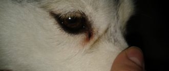
The dog squints its eye - what happened?
The condition of a dog's eyes can tell a lot to an attentive owner. If the eyes are clean, clear and shiny, without redness, swelling or excess discharge, this indicates that everything is in order and the pet is healthy. Any deviation from the norm should alert the owner.
A healthy dog's eyes are always clean and clear.
To some, due to inexperience, it may seem funny how a dog grimaces, squinting one or both eyes, but there is absolutely nothing funny about this - in this way the animal gives the first signal of its ill health.
Possible reasons
Squinting of the eye can be caused by one of the following reasons:
- erosions and ulcers of the cornea caused by trauma;
- foreign objects entering the eye;
- rubbing with hair and eyelashes;
- viral infections;
- chlamydia;
- eye burn;
- acute purulent conjunctivitis;
- allergies;
- dry eye syndrome;
- glaucoma, etc.
Eye diseases pose a serious risk to your pet. If the most seemingly harmless disease at first is not cured in a timely manner, its course can become more complicated and lead to loss of vision or even the death of the animal.
Associated symptoms
Most often, eye diseases have complex symptoms, and you need to pay attention to not just one, but several manifestations of the pathology; the same applies to cases of eye injury. The difficulty is that similar symptoms can characterize various ophthalmological problems, and the problem of self-diagnosis leads to error and aggravation of the disease.
Heavy discharge from the eyes
Excessive, bad-smelling, and especially purulent discharge is not normal . To determine and eliminate their cause, you should immediately consult a doctor.
Discharge from a dog's eye can also be caused by non-ophthalmological reasons - for example, worms
Video: why your pet's eyes may become watery
Swelling and redness
Typically, excessive discharge from the eyes is accompanied by the following phenomena: redness of the whites of the eyes and the epithelium of the eyelids, the area around the eye socket becomes swollen, and itching may occur.
Temperature increase
A temperature of up to 39 degrees is considered normal for dogs; in small breeds this figure may be slightly higher. An increase in temperature is clear evidence that an infection has occurred and an inflammatory process is developing. This is a very alarming symptom, which is often combined with refusal to eat and general weakness.
Photophobia
Fear of light is one of the characteristic signs of acute conjunctivitis, a dangerous infectious eye disease. The eyeball is very inflamed and painful, it is difficult for the dog to look at the light and especially at the bright sun - the animal experiences a sharp pain and tries either not to open its eyes at all or to squint them too much.
If your dog has conjunctivitis, it hurts to look at bright light.
Diagnostics
Only a veterinarian can make an accurate diagnosis; this may require special equipment or tests, which can only be done in a clinic. But the owner himself can carry out a primary, albeit very approximate diagnosis, while observing the rules of caution:
- Wash your hands well with soap and dry them with a clean towel.
- Bring and lead the animal to a source of bright light - a window or lamp.
- Calm your dog and very carefully examine the inside of each eye and eyelids.
Your first task is to rule out or confirm eye injury. Further actions will depend on the results of the initial examination. Try to examine the eye from all sides and with side lighting - the intact cornea will look smooth and intact, and the foreign body will become clearly visible.
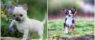
Tearing common to certain dog breeds
Tearing is normal in some breeds. For example, brachycephalic dogs, long-haired dogs, as well as in breeds with an enlarged palpebral fissure, the secretion of tear fluid will be normal. These breeds require regular care and careful eye treatment. Breeds with this feature include:
- Saint Bernard;
- bloodhound;
- mastiff;
- Shar Pei:
- cocker spaniel;
- bullmastiff;
- bulldog;
- chow-chow;
- Basseta;
- decorative dogs (Yorkshire terrier, Pekingese, etc.).
Bulldogs are prone to lacrimation
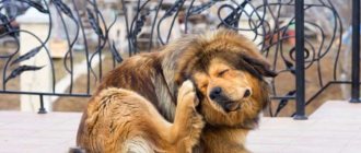
What should you do if your dog’s ears, eyes, muzzle, paws or other parts of the body are constantly itching?
- If lice, fleas or lice eaters are found, the dog, as well as the floor in the enclosure or apartment, must be treated with a solution of neostomozan, butox, and so on.
- After treatment, 5 days later you need to put a special anti-flea collar on your dog.
- If you suspect linguatulosis, otodecosis, sarcaptosis, or cheyletiosis, it is better to consult a veterinarian for an accurate diagnosis and a correct diagnosis.
- You cannot do without a specialist if you identify an allergen or suspect demodicosis.
Dog eye examination
It is necessary to regularly examine your pet, and do not forget to pay attention to the eyes. A large dog can be examined by placing it on the floor, and a small dog can be examined on a table by sitting or laying it down. Carefully, without effort or pressure, use the fingers of your free hand to slightly open the palpebral fissure. This will allow you to examine your dog's eyes for the presence of a foreign body or determine the type of discharge from the eye.
Examination of the organs of vision must be done very carefully so as not to harm the pet.
What you need to know! If the dog does not allow you to approach him or shows aggression, trying to defend himself, then you need to carefully secure the mouth with a wide braid. In this case, a muzzle may make inspection difficult.
A healthy animal is distinguished by clarity, cleanliness, with the absence of excessive eye fluid, as well as the presence of a transparent cornea of the eye. The outer membrane of the eye (the tunica albuginea) is white, without inclusions or bloody stains. If there is a characteristic change in the eye, then this indicates the spread of the pathology.
Regular inspection is carried out in a well-lit place, preferably in the morning and after walking. It is necessary to examine the condition of the eyeball as a whole, the conjunctiva, the absence of redness, swelling, the absence of a foreign body, the normal position of the organs of vision, the reaction of the pupil, the absence of various types of formations.
Every year, take your dog to see an ophthalmologist to examine the condition of the cornea and its internal structure, and to rule out the presence of ophthalmological pathology. If the cornea has changed color, it may be an ulcer or injury. Deformation of visual functions, blepharospasm, lacrimation, and the appearance of a nictitating membrane are symptoms of an ophthalmological disease. Yellowing of the conjunctiva indicates a problem with the liver, possible hepatitis, helminthiasis. Blue discoloration is, in turn, a symptom of intoxication poisoning in a dog.
The dog must be taken to an ophthalmologist annually
For preventive purposes, you can wipe your pet's eyes with a strong tea infusion without additives and chamomile infusion.
Treatment of eye diseases in dogs, medications for eye treatment in a veterinary kit
Treatment of conjunctivitis
When treating conjunctivitis, on the recommendation of a veterinary specialist, the owner can use these drugs:
- antibacterial drops ("Tsiprovet" - 1-2 drops/4 r per day for 1-2 weeks; "Bars" - 1-2 drops/4 r per day; "Anandin" - 2 drops/2 r per day);
- antibacterial ointment (“Conjunctivin” - 2-3 drops/3 times per day for 5-10 days).
Eye drops "Tsiprovet"
Treatment of an injured eye
If an eye injury is detected in a dog, first aid is necessary. When examining the eye and detecting a foreign body (splinter, grass, fragments, pebbles), it is necessary to rinse the eye with an anesthetic (Oxybuprocaine hydrochloride, Benoxy - 1-4 drops), and only then proceed to remove the object. With such an intervention, do not forget about the use of antibacterial drugs.
Before starting treatment, you need to visit a veterinary clinic, only there they will examine the dog and prescribe the correct treatment. The specialist will first prescribe means for washing the eyes from purulent accumulations, and will tell you what materials to use to treat the eyes. The next step is to prescribe eye drops, which are used strictly after washing the eye from pus. Antibacterial and anti-inflammatory drugs are prescribed for infectious diseases. Vitamins can also be prescribed to strengthen the immune system. During the treatment period, special socks and a collar may be used to limit access to diseased areas.
What you need to know! Try to strictly adhere to the specialist’s instructions, especially with regard to the dosage of eye drops, since dogs have very developed sensitivity of the visual organs.
It is necessary to instill medications according to the veterinarian's prescription.
In case of eye injury, first aid is provided, so the first aid kit should always have painkillers (1-2% novocaine solution) for disinfection (1% brilliant green solution). The treated surface is covered with a sterile bandage, and the dog is taken to a veterinary clinic.
Treatment of blepharitis
Inflammation of the eyelid (blepharitis). Usually provoked by careless scratching, a tick bite, or a bruise. Bleeding ulcers at the base of the eyelashes, with the appearance of dried crusts and scales, at the same time there is thickening and redness of the eyelids, loss of eyelashes, severe lacrimation - a manifestation of blepharitis.
The following drugs are used for treatment:
- "Sofradex" (strictly as prescribed by the doctor);
- “Lakrikan” (1-2 drops 2-3 times a day for 8-10 days);
- "Calendula" 10% ointment for external use;
- “Hydrocortisone acetate” (0.2 ml 2-3 times a day).
Video - How to put drops in a dog's eyes
Treatment of inflammation of the conjunctiva
Inflammation of the conjunctiva. It occurs due to a deficiency of various vitamins and minerals in the dog’s body, or the penetration of a foreign body.
The following drugs are used for treatment:
- "Bonkharen" (1-2 drops every 2-12 hours for 5-7 days);
- “Veteritsin gel” (3-4 times a day);
- “Desacid” (2-4 drops 3-4 times a day for 7-10 days);
- “Dekta-2” (2-3 drops 2-3 times a day for 5-10 days);
Eye drops "Desacid"
Treatment of keratitis
Keratitis. Inflammation causes clouding of the cornea of the eye, which, without proper treatment, can lead to perforation of the cornea itself and cause the appearance of ulcers and abscesses. Inflammation of the cornea can be superficially purulent, deeply purulent, superficially vascular, catarrhal, phlyctenulous, punctate, and superficial.
Depending on the type of inflammation, medications are used:
- "Lacrimin" (3-4 drops daily);
- “Forvet” (1 drop 4-5 times a day for 7-30 days);
- "Sofradex" (strictly as prescribed by the doctor).