Why does this happen?
The cause of uterine prolapse in dogs is usually a difficult birth with heavy blood loss. Due to multiple pregnancy, stretching of the reproductive organ may occur. Forcibly removing a puppy during labor provokes prolapse or prolapse of the uterus.
What else can provoke the disease? Common reasons:
- Obesity or wasting.
- Poor nutrition.
- Mineral fasting.
- Lack of proper walks.
- Childbirth in old age.
- Old age.
During pregnancy, complete loss of a reproductive organ rarely occurs. At first, periodic protrusion of the vagina may be seen. If you do not catch it in time, the disease may drag on. An unpleasant odor from the discharge from the animal’s genitals, purulent discharge, difficulty urinating - this is a wake-up call for the owner. You should not delay visiting the veterinarian.
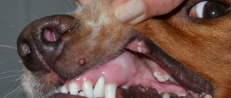
Symptoms
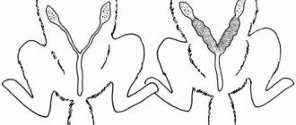
Loss can be total or partial. With complete prolapse, the bicornuate red uterus hangs almost to the ground. In case of partial loss, a piece of pink tissue protrudes from the loop, then hides again. However, if there is no attention to the problem, the size of the protruding tissues increases during the next heat or for another reason. The dynamics of the pathological process are characterized by the following signs:
- the bitch constantly looks around, trying to see what is happening to her from behind;
- the animal loses activity and stops eating;
- licks the vulva;
- intermittent fever develops;
- the mucous membranes of the gums and lips become white: blood rushes to the reproductive organs;
- the organ leaves the loop and enters again;
- the loss becomes permanent, the mucous membrane dries out, cracks, and foci of necrosis appear;
- the range of colors varies from bright red to purple;
- a specific smell appears.
The success of treatment depends on the timeliness of seeking veterinary help. The appearance of an odor and purple color are symptoms that threaten the life of the animal.
How to help?
When uterine prolapse occurs in dogs, what should owners do? Provide your pet with the necessary assistance before the veterinarian arrives.
The female is placed on her side so that her butt is slightly raised. The uterus should be wrapped in a clean cloth. This is done in order to prevent contamination of the organ and, as a result, the occurrence of cracks. If dirt gets on the fallen part, it is carefully removed with a damp cotton swab.
Symptoms of uterine prolapse
It is not so difficult to notice that an animal has a serious gynecological problem. The owner pays attention to the following symptoms:
- Anxiety of the animal, forced posture, frequent pushing.
- The dog constantly licks the perineal area.
- From the genital slit there is a protrusion of a cylindrical organ, the apex of which is depressed (uterine horn).
- The mucous membrane of the prolapsed organ initially has a pink-red color and bleeds. As the uterus is outside the animal's body, its color changes to dark red, then blue and purple. Swelling of the mucous membrane occurs, the uterus increases in size.
- As the mucous membrane dries, cracks and ulcerations form on the organ. In severe cases, tissue necrosis develops.
Treatment
What is the treatment for uterine prolapse in a dog? Either returning it to its original position, or amputating the reproductive organ. The animal must be hospitalized. And then the doctor makes a decision on the situation. If necrosis has begun or the soft tissues are very contaminated, the veterinarian will amputate the organ. If the tissue is viable, the uterus is returned to its place through surgery. For uterine prolapse in dogs, the operation is performed under anesthesia. This is a must.
The sooner the animal receives the help it needs, the better. If it is not provided on time, the pet will die from blood poisoning.
If everything is not so scary, then the doctor bandages the base of the fallen part. After 6-10 days it disappears. The dog is under the supervision of a specialist all this time.
When it is possible to realign the reproductive organ, if it is viable, the uterus is treated with detergents and realigned.
Vaginal prolapse in dogs
Vaginal prolapse (vaginal prolapse) is characterized by complete or incomplete eversion of the vaginal walls through the genital slit to the outside.
This disease in dogs usually occurs during estrus and is associated with increased secretion of hormones into the blood; very rarely, vaginal prolapse occurs during pregnancy. Vaginal prolapse is most common in St. Bernards and Boxers.
Etiology . Vaginal prolapse in a dog occurs as a result of relaxation of the paravaginal tissue that secures the vagina, often due to increased intra-abdominal pressure. Increased secretion of hormones during estrus. Inadequate feeding, exhaustion, obesity, mineral starvation, hypovitamin deficiency, keeping a dog without walking outside, multiple pregnancies, difficult childbirth, old age, etc. predispose to vaginal prolapse.
Clinical picture . In dogs, there is usually only partial vaginal eversion of varying degrees. Complete vaginal prolapse is rare in dogs during pregnancy. At the onset of the disease, the dog owner notices periodically appearing protrusion of the vagina, especially during the act of urination, or can be repeated with each emptying. The dog is worried and licks the prolapsed vagina. During clinical examination, a spherical fold of pink-red color appears from the swollen loop. The mucous membrane of the prolapsed vagina swells, becomes dirty, injured, becomes tight and rigid. If the disease drags on, we notice purulent discharge from the vulva and vagina, the dog has difficulty urinating, and foul-smelling discharge appears from the external genitalia. During a clinical examination, we detect ulcerations, vesicles, papules, pustules, erosions, and necrosis on the vagina.
The diagnosis is made based on the clinical symptoms of the disease.
Differential diagnosis . When diagnosing vaginal prolapse, the veterinarian excludes this disease from tumors of the vaginal wall (leukomyoma and venereal sarcoma). Vaginal swelling is common in young female dogs during proestrus or oestrus.
Treatment .
In case of incomplete vaginal prolapse, the base of the prolapsed part of the vagina is tied with an elastic band. As a result of this, the bandaged vaginal tissue dies and disappears after 6-10 days. The bladder is first catheterized. Under no circumstances should the tissue be cut off after ligation. bleeding occurs. In case of complete vaginal prolapse, it is treated with detergents (10-20% solution of dimecaid, 0.1% solution of miramistin, use citeal, etonium, decamethoxin) and set. If there is partial vaginal prolapse and signs of labor appear, a cesarean section is necessary. In those rare cases where the vagina prolapses completely, realignment usually requires widening the vulva through a perineal incision (perineotomy). Having straightened the vagina, it is strengthened by applying a circular suture at the border of the vagina and the vestibule, taking care not to damage the urethra. After this, the perineal wound is sutured with two rows of stitches: from the mucous membrane - with catgut, from the skin - with silk. Before birth, the applied circular suture is removed.
Prevention
What measures should be taken to prevent uterine prolapse in a dog?
- Firstly, it is nutritious nutrition throughout the dog’s life. This is especially true during pregnancy.
- Secondly, vitamin complexes must be present in the animal’s diet.
- Thirdly, you need to walk your pet for more than five minutes twice a day. The walking time depends on the needs of the breed. At least 20 minutes for small breeds and 40 minutes for large breeds.
- The fourth item is dog bed. The animal's sleeping place must be clean and located on a flat surface.
- Fifth, examination by a veterinarian during pregnancy. It is advisable to perform an ultrasound to detect multiple pregnancy.
- The sixth moment is childbirth. During childbirth, the presence of a veterinarian and the owner is required with the dog.
- The seventh point is to carefully monitor the dog in the first 12 hours after the birth of the puppies. Uterine prolapse most often occurs during this time period.
- If the owner sees that the pet is in trouble, immediately call a veterinarian.
Causes of uterine prolapse in dogs
In obstetric practice, it is customary to distinguish between the causes of uterine prolapse in the postpartum period and factors unrelated to pregnancy and labor in dogs.
After childbirth
Most often, the disease is diagnosed during childbirth after the birth of the penultimate or last newborn. The risk group includes animals that have given birth a lot, as well as dogs of small breeds. The causes of uterine prolapse during this physiological period, veterinary experts include the following:
- Multiple births. The presence of a large number of fetuses in the womb leads to severe overstretching of the muscular layer of the uterus. This phenomenon leads to a decrease in the tone of the reproductive organ and provokes its prolapse beyond the anatomical boundaries.
- Hydrops of the fetus and membranes. A pathological increase in the size of the future puppy is a common cause of prolapse or inversion of the uterus due to relaxation of the ligamentous apparatus.
- Violent labor due to hormonal overload of the body.
- Illiterate obstetric care. Forced extraction of the fetus when the birth canal is dry is often accompanied by obstetric pathology.
- Heavy bleeding during labor.
Veterinary experts include illiterate feeding of the pregnant female as factors that provoke complete or partial prolapse of the uterus in the postpartum period. A low level of protein in the diet, a large proportion of bulky feed, frequent stress, and unsatisfactory living conditions are significant reasons for the development of the disease.
The lack of physical activity of a female during pregnancy and obesity, according to experts, lead to a decrease in the tone of the smooth muscles of the reproductive organ and provoke obstetric pathology in the postpartum period.
In nulliparous
Uterine prolapse in nulliparous dogs is an extremely rare phenomenon and is usually caused by injuries to the abdominal organs and the presence of neoplasms in the womb. Most often, the owner is faced with such a phenomenon as vaginal prolapse - protrusion or complete prolapse of the organ from the genital slit to the outside, and may mistakenly mistake this phenomenon for uterine prolapse.
During heat
In addition to the postpartum period, the dog owner may encounter such a phenomenon as protrusion of the reproductive organ outside the body during estrus. Boxers and bulldogs are predisposed to the pathology.
The cause of the disease is a hormonal imbalance. Under the influence of estrogens, hyperplasia occurs in the uterus and vagina - thickening of the walls of organs. This leads to disruption of the muscle tone of the womb and contributes to disruption of the anatomical position of the reproductive organ, up to eversion outside the body.
Let's summarize
Let us highlight the main aspects of the article:
- Uterine prolapse is more common in dogs that repeatedly give birth.
- Representatives of small breeds are prone to the disease.
- Loss occurs within 12 hours after birth.
- It can be provoked by poor living conditions: lack of adequate walks, lack of vitamins and poor nutrition.
- It is extremely rare, but uterine prolapse can occur during estrus. This is due to the hormonal levels of the animal.
- Treatment is possible in several ways: amputation, reduction and ligation of the prolapsed part.
- After surgery, you must strictly follow all doctor's recommendations.
Causes of uterine prolapse
The main factors leading to uterine prolapse: difficult or improperly performed childbirth and everything associated with this process - severe physical stress, heavy bleeding.
This can also lead to:
stretching of the reproductive organ;
overstretching of membranes;
poor nutrition of the female during pregnancy.
Forcible removal of the fetus can also contribute to eversion of the dog's reproductive organ. In some cases, prolapse is provoked by estrus.
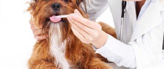
Diagnosis of an animal
Having discovered prolapse of the vagina or uterus in his pet, the owner should take measures for an early visit to a veterinary specialist. Only a qualified doctor can make the correct diagnosis based on anamnestic data, examination, palpation, vaginoscopy, and laboratory tests.
A clinical blood test helps determine the general condition of a sick animal. A number of manipulations, such as vaginoscopy, are performed under anesthesia. If necessary, cytological examination of tissues is performed.
First aid and treatment
Let us warn you right away - you should not try to straighten the uterus without the appropriate skills and necessary tools! This will only make the owner worse. Instead, the following measures should be taken:
- The dog is placed on its side, the organ is thoroughly washed with a solution of furatsilin, miramistin or chlorhexidine.
- The uterus is wrapped in sterile gauze wipes, which are also well moistened with any of the above solutions.
After this, the animal is immediately taken to the veterinarian. It is not worth doing the reduction at home, as this is a rather complicated procedure. In addition, unfortunately, it is possible that the uterus will have to be removed.
Care and prevention
Caring for an animal after repositioning the reproductive organ comes down to following the veterinarian’s recommendations. If surgical removal was performed, the postoperative period is reduced to treating the sutures with antiseptic drugs and systemic use of antibacterial agents.
Prevention of uterine prolapse in dogs is possible if the owner follows the following rules and recommendations of veterinary specialists:
- Balanced feeding of a pregnant female eliminates many gynecological problems in the postpartum period, including uterine prolapse.
- Regular dosed physical activity during pregnancy allows you to maintain the smooth muscles of the reproductive organ in good shape.
- Providing qualified assistance to an animal during childbirth.
Partial or complete uterine prolapse is most often diagnosed in dogs immediately after birth. In rare cases, pathology can be observed during estrus. A sick animal needs qualified help. In some cases, a veterinarian performs an ovariohysterectomy to save the dog’s life. In uncomplicated variants of the disease, reduction of the prolapsed organ is used with further antibacterial therapy.
Causes of the disease
For parous and nulliparous dogs, the causes of uterine prolapse vary greatly. Let's first consider the main factors for animals that have not had puppies:
| Injuries | Injuries and bruises in the pelvic organs. This often happens when struck by a person or in a collision with a car. |
| Hormones | Any hormonal therapy can weaken the uterine tissue. Background disturbances due to natural causes are also possible. |
| Heredity | Predisposition due to heredity. If the bitch suffered from this disease, then the puppy will also inherit this pathology with a probability of 40-50%. |
| Chronic inflammation | The presence of chronic inflammation. Loss is almost always accompanied by pyometra. |
| Congenital pathologies | Organs of the genitourinary system, as well as defects acquired as a result of improper conditions of detention. |
| Age | Age-related changes provoked by a general weakening of tissues due to poor nutrition. |
| Bleeding | The presence of constant internal bleeding from the capillaries. They can only be detected by ultrasound. Erosion processes gradually corrode the mesenteries that hold the uterus in place. The initial condition may be infection or heatstroke. |
In animals that have given birth, the catalyst is usually estrus. The action of sex hormones negatively affects the general condition of the animal, weakens it, and prepares the environment for conception. If the reproductive system is not firmly in place, then all these changes can lead to protrusion through the vagina. Loss can also occur due to parasites in the body, constant stress if the owner overtrains the animal for exhibitions or hunting, poor hygiene, viruses and bacteria.
Factors provoking the development of pathology
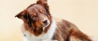
Adenoma of the third eyelid in dogs does not have the specificity of spreading to the eyeball itself. But the disease should not be written off. This is due to the fact that the disease does not go away on its own and the symptoms do not disappear.
If the owner notices a hypertrophied pinkish bulge in the inner corner of the dog’s eye, it is important to contact a qualified veterinarian. Pathological changes that develop in the inner part of the corner of the eye, caused by a benign neoplasm, become factors of additional discomfort in the dog.
The treatment method used is surgical excision.
After surgery to remove the affected area, the animal suffers from discomfort associated with the development of dry keratitis and inflammation of the conjunctival membrane.
There is a certain group of purebred dogs with a genetic predisposition to the development of third eyelid adenoma. These include: pugs, spaniels, Mexican and Chinese Cresteds. A neoplasm is diagnosed at the location of the third eyelid in breeds such as German Shepherd, Doberman, Pinscher and Great Dane.
Other dog breeds also suffer from the pathological process, but much less frequently. The basic factors that provoke the disease are:
- increased weakness of the ligamentous apparatus of the eyeball (their main function is to maintain the lacrimal gland in a normal physiological position);
- natural phenomena in the cartilage of the eyelid;
- the occurrence of complications after leukemia in the form of hyperplasia;
- factors of genetic inheritance;
- injuries sustained by the dog as a result of active games, fights with relatives, or scratching with claws;
- processes of a pathological nature developing in the eyeball itself, provoking increased secretion of the lacrimal glands in the area of the third eyelid.
Treatment of uterine prolapse in dogs
Before receiving qualified assistance, the animal owner must be able to provide first aid. First of all, you need to place the dog on a clean bedding. Using sterile wipes, pre-moistened with a cold disinfectant solution, wrap the prolapsed organ. A solution of chlorhexidine, potassium permanganate, furatsilin, Miramistin is used as an antiseptic.
After the animal is diagnosed, the veterinarian, based on the severity of the obstetric and gynecological pathology, will choose the best way to solve the problem.
If the organ has not completely prolapsed and the outer part does not show signs of inflammation, then in some cases they can manage by repositioning the uterus. The tissues are treated with cool antiseptic solutions, and if necessary, a novocaine blockade with an antibiotic is carried out. After irrigation with disinfectant solutions, the uterus is sprinkled with streptocide powder.
Reduction of the uterus followed by sterilization
Manipulation to return the organ to its anatomical position is carried out using anesthesia, for example, under a sacral blockade. To return the uterus to the abdominal cavity, special nylon sticks are used. To prevent the development of inflammation, a sick dog is prescribed broad-spectrum antibacterial drugs, usually cephalosporins. After reduction, the veterinarian applies special loop-shaped sutures to the vulva.
Sutures for holding the vagina: a) loop-shaped; b) roller
If serious tissue damage has occurred and there is a risk of organ necrosis, a veterinary specialist will perform amputation of the uterus. For animals that are not of breed value, ovariohysterectomy is recommended. Sterilization of the dog is also necessary if uterine prolapse is regular.
For information on surgical reduction of the uterus in dogs, watch this video: