Our four-legged friends, unfortunately, are often susceptible to various injuries: including possible injuries to the ears. Many owners are familiar with such a pathology as otohematoma in dogs. In the article we will look at the features of this injury, find out why otohematoma appears, how to treat it, and what to do to prevent this pathology.
Are you here
Description and reasons
Otohematoma is an accumulation of fluid or blood in the wall of the auricle due to rupture of lymphatic and/or blood vessels.
Otohematoma forms on the inside of the auricle - between the skin and cartilage. The mechanism of hematoma development is as follows: when blood vessels rupture on the inner surface of the vessel, blood or lymph under pressure peels off the skin from the underlying cartilage. The formation of a cavity of fluid under pressure causes pain to the animal; he reacts to it by scratching with his paw or rubbing his head, which further increases the pressure in the hematoma cavity and leads to its further increase.
The exact mechanism of primary vascular damage is not fully determined. According to one theory, it develops with mechanical damage to blood vessels, during events such as shaking the head and scratching with the paws (more often, with inflammation of the outer ear). But quite often at an appointment at a veterinary clinic, the exact cause of the development of otohematoma cannot be detected. In such cases, veterinarians suggest various allergic and autoimmune diseases as the cause, but definitive evidence has not yet been obtained.
Clinical signs
Otohematoma develops in both cats and dogs, most often forming on one side, in rare cases both ears are involved in the process. Externally, otohematoma has characteristic manifestations; upon examination at a veterinary clinic, it is very difficult to confuse it with another disease.
At the initial stage of hematoma formation, it can be small in size, but can be easily felt by the veterinarian as a dense, fluctuating swelling. In the absence of treatment, its size increases significantly, and the affected area can reach the entire surface of the outer ear. In chronic cases, the liquid contents of the otohematoma thicken and become rigid; at this stage, deformation of the auricle may be observed. Sometimes, an otohematoma can spontaneously burst, revealing an accumulation of fluid inside the ear and signs of inflammation.
As differential diagnoses of otohematoma, only two diseases are considered - neoplasms (tumors) of the external auditory canal and cysts. To exclude these diseases and make a final diagnosis, a doctor at a veterinary clinic can undertake some diagnostic tests - puncture the cavity to obtain fluid (puncture), examine this fluid under a microscope (cytology), conduct an ultrasound examination of the cavity, examine the inner ear with an otoscope and collect material for subsequent cytological examination (identification of mites of the external auditory canal and the type of microflora).
It should be remembered that even if the animal does not show much concern, otohematoma is quite painful for the animal, and most of its treatment should be carried out under anesthesia.
Treatment
When a hematoma occurs, there are both conservative (medicinal) and operative (surgical) treatment methods. Conservative treatment methods include drugs such as corticosteroids (eg prednisolone, dexamethasone) and antibiotics (eg cefazolin), but it should be remembered that otohematoma is a surgical disease, the best results in its treatment are achievable only by using surgical methods.
If fluid accumulates, it must be evacuated as soon as possible (at the time of contacting the veterinary clinic), otherwise the risk of increasing the affected area increases significantly. In the future, various techniques are used to constantly drain fluid and increase the rate of fusion of the cavity walls. It is the use of surgical correction methods that leads to the fastest resolution of the process and most completely prevents ear deformation.
In our veterinary clinic, the main type of surgical treatment is dissection of the otohematoma cavity with a scalpel, followed by suturing the entire thickness of the ear with nylon sutures. The animal is under anesthesia, a skin incision is made on the inside to drain fluid, the walls of the cavity are sutured with interrupted sutures over the entire area of the lesion with the node located inside. Sutures are applied in a staggered manner to create better conditions for resolving the cavity and reducing deformation of the external auditory canal. Immediately after the operation, some fluid discharge is noted from the skin incision, which within a few days significantly decreases and disappears. Nylon sutures do not cause any tissue irritation and can remain in place for a very long time, but it is better to remove them 14-21 days after the operation. While the sutures are in place, scar tissue forms in the area where they are located, which closely fuses the skin with the underlying cartilage of the ear. Recurrence of otohematoma after surgical treatment is possible, but unlikely.
As surgical treatment options, other techniques can be used - the placement of various types of negative pressure drains, laser excision of the walls, etc. But, in our veterinary clinic we use exactly this technique, it has been used for years, it suits both cat and dog owners and veterinarians; it doesn’t make much sense to look for other methods of correction.
At the time of treatment of otohematoma, attempts are also made to identify underlying diseases, usually inflammation of the outer ear. Depending on the results of the examination, the animal owner may be recommended to carry out various forms of treatment for external otitis, this is done to reduce the risk of relapses.
Anesthesia
Separately, it is worth mentioning about anesthesia; correction of otohematoma is possible only under anesthesia, but every anesthesia treatment carries a potential anesthesia risk, including a fatal one. Before administering anesthesia, the doctor at the veterinary clinic conducts a physical examination of the animal; if any abnormalities are detected, he recommends diagnostic tests and selects optimal protocols. But no matter how hard the clinic’s veterinarian tries, he can kill your animal with any injection.
Causes of lymphoextrovasation in cats and dogs
In most animals, auricular hematoma develops as a result of excessive head shaking or chronic, persistent scratching and damage to the ear. This can occur immediately after bathing the animal, due to chronic otitis media or an allergic reaction, which causes severe itching and inflammation. The presence of parasites (for example, demodex mites) or a foreign object in the auricle can also indirectly affect this pathological process.
Shaking your head can cause these tiny blood or lymph vessels to rupture, bursting and spilling into the surrounding area, gradually peeling the skin away from the cartilage tissue. The accumulated exudate causes excessive irritation, and the animal begins to shake its head even more, thereby aggravating the process. If the situation is not resolved, then blood or lymph continues to accumulate under the skin in greater and greater quantities, increasingly peeling the skin away from the cartilage and forming a large pocket, which in some cases can block the ear canal.
In some cases, constant shaking of the head can eventually lead to spontaneous rupture of the hematoma and splashing of its contents outward. However, a responsible pet owner should never allow such a development to occur.
Pathogenesis of the disease
After the vessel ruptures, blood flows out and permeates the surrounding tissue. The pressure of the flowing blood pushes and separates the tissues, which leads to the formation of a pathological cavity (hematoma). The process of bleeding stops when the pressure inside the vessel is not balanced by the resistance of the walls of the created cavity.
Therefore, hematomas with arterial injuries are always larger than with veins. In addition, the size of the hematoma is determined by blood pressure, the type and extensibility of the skin, and blood clotting.
When hematomas form, a process of self-blocking of hemorrhage occurs in parallel. First, the ends of the damaged vessels curl up and are drawn into the surrounding tissue. At the same time, the lumen of the vessels narrows. Thanks to this, a strong blood clot forms within 2-3 days.
The process of self-blocking hemorrhage does not occur if there is only a crack or puncture near the surface of the artery. In this case, a parietal pulsating hematoma is formed, and then a false aneurysm is created from it.
In the created cavity, the blood coagulates, a small thrombus resolves on its own, while in case of a large hematoma it remains for life. In this case, the blood clot is replaced by connective tissue, then a capsule is formed, in which calcium salts are later deposited, resulting in a cyst. When microorganisms penetrate, an abscess or phlegmon occurs.
Lymphoextravasate in dogs: main symptoms and treatment
Lymphatic extravasation in dogs usually occurs due to traumatic exposure to heavy objects, bruises and falls . The skin is not damaged, but a cavity is formed under the skin in which lymph from damaged lymphatic vessels is located. Dogs often experience lymphatic extravasation of the ear , which is caused by excessive irritation and shaking of the dog's ears. The skin of the ears is very delicate, and the blood and lymphatic vessels that pass here are also thin and fragile, which is why they are damaged with the outpouring of lymph, and sometimes blood, under the skin. This leads to its detachment from the ear cartilage and the appearance of a cavity filled with fluid .
Treatment of ear with hematoma
Treatment of ear hematoma in a dog is carried out in two stages. On the first, the main task is to stop hemorrhage and hematoma formation. It proceeds to the next stage only after the formation of a durable blood clot. At the second stage, measures are taken to quickly resolve or remove clotted blood clots.
First stage
This help can be provided at home. The skin is treated with antiseptics, and a cold compress is applied to the hematoma area: heating pads and bags of ice, cold liquid, and cooled meat.
The procedure must be combined with the creation of a pressure bandage:
- the ear is pressed to the head, since it will not be possible to apply a bandage to one ear;
- bandage the entire head, like a scarf;
- The bandage must be firmly in place, because the dog will probably try to remove it.
Before applying the bandage, you can use a sterile needle to remove the blood, and also introduce novocaine-antibiotic solution to eliminate pain and prevent purulent processes.
Second phase
Treatment of minor hemorrhages
Small hematomas (up to 1 cm in diameter) resolve on their own. To increase the speed of the process, thermal procedures are carried out: heating the hematoma area with a Sollux lamp, vaporization, application of ozokerite, paraffin. Resorbing ointments are also prescribed. But for large hematomas, the above actions are not effective.
Treatment of medium and large hematomas
2-3 days after the injury, blood begins to clot; the blood clot reaches its maximum strength on days 4-5.
At this time, the hematoma should be opened or its contents sucked out. At the same time, blood clots cannot be torn off by force; such damage to the protective function of the clot will lead to repeated hemorrhage.
After removing the blood clot, the wound is disinfected with a 0.02% solution of furatsilin, then powdered with Platokhin’s compound antibacterial powder. At the end the wound is sutured. Incomplete suturing is acceptable if it is necessary to ensure blood flow. In case of purulent processes, the wound is not sutured at all.
The exfoliated skin must be sutured to the underlying tissues (perichondrium) using a stitched roller suture. After the operation, a pressure bandage is again created so that the walls of the hematoma come closer together without tissue resistance. The stitches are cut off after 10 days.
For a pulsating hematoma, surgery is performed before a blood clot forms (on the day of treatment). They dissect the tissue, find the bleeding vessel, and use their fingers to close the vessel upstream. Then the vessel is sutured using a ligature. Then the wound is drained, freeing it from blood and its clots, after which it is treated with antiseptics and sutured.
Blood pumping technique
A more conservative method of therapy without surgery.
The hematoma is removed with a syringe.
- The dog is securely restrained, as sudden movement can lead to additional injury. Anesthesia is not required as the treatment is minimally invasive.
- The surgical field is prepared. A desirable, but optional procedure, since the dog’s skin is highly resistant to surrounding microorganisms.
- A puncture is made in the upper edge of the ear, the blood is sucked out as completely as possible, but if a small amount remains, it will later resolve itself.
- After sucking out the blood, the needle is not removed, but another syringe is connected and a mixture of antibiotics and novocaine is injected. The amount may vary and depends on the characteristics of the disease, the condition of the pet and the decision of the veterinarian.
- At the end, the ear is tightly bandaged with a pressure bandage. A collar is attached to prevent the animal from scratching its ear.
Treatment of lymphatic extravasation in dogs
The prognosis for this disease is almost always favorable, except in cases where it is complicated by infection of the lymph by pathogenic microbes.
Different types of disease require different approaches to treatment, and sometimes surgery is necessary. This is especially true for lymphatic extravasation of the dog’s ear, since its conservative treatment by puncturing the cavity and suctioning fluid from it often leads to re-filling of the “pocket” with lymph literally in a day or two.
Conservative treatment of lymphoextravasate in dogs
The main requirement in caring for a sick dog is as much rest as possible, since lymph circulation during active exercise increases fivefold, and with this disease it is important, on the contrary, to reduce its secretion and help the body create blood clots in injured lymphatic vessels. Neither applying cold objects nor warming the cavity is indicated , as they will only cause side effects in the form of possible tissue necrosis or increased lymph secretion. Massage that causes increased movement of blood and lymph through the vessels also contraindicated
If the lymphatic extravasate is superficial and occupies a small area , then the veterinarian will choose conservative-operative treatment tactics. On the first day, use pressure, wet compresses with various alcohol solutions. Punctures are also used to empty the cavity of exudate and add a 1-2% iodine solution to it, after which a pressure bandage is applied. Such manipulations are performed more than once. and the course. If this treatment is ineffective, then surgical methods - an operation to empty the cavity of fluid and apply sutures to speed up its healing.
Reasons and method of surgical treatment of lymphatic extravasation in dogs
The surgeon's intervention implies careful adherence to the rules of antisepsis and asepsis, since it is important to prevent the introduction of pathogenic microflora into the cavity. To do this, the skin at the bottom of the cavity is cut with a small incision, the contents are removed, and a drainage soaked in antiseptic solutions is inserted into the cavity. After two days, the drainage is removed, treating the incision site with Vishnevsky ointment and sulfonamides. Such measures will allow the dog to recover 14-20 days after surgery , a maximum of a month.
In the surgical treatment of ear lymphatic extravasation in dogs, different surgical techniques are used. Their essence also consists in removing fluid and applying several sutures - stitches along the surface of the auricle so that the skin and ear cartilage grow together, and the damaged vessels stop releasing lymph or blood. The stitches will not be removed for about three weeks. During this time, scars appear, which are necessary for the healing of damaged tissues and prevent relapses of lymphatic extravasation.
Otohematoma - what is it?
Otohematoma - what is it? - (auricular hematoma) is an accumulation of blood and lymph on the inside of the auricle, between the cartilage and the skin.
The main cause of otohematoma is traumatization of the vessels located on the auricle. As a rule, these injuries are self-induced (i.e., inflicted by the animal on itself), as a result of itching (caused by otitis media of various natures, flea dermatitis, and also insect bites). There is also a scientific version of the immune-related origin of hematomas.
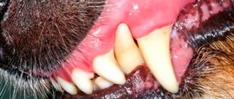
At the site of otohematoma formation, a limited swelling of a round or oval shape forms. The hematoma is usually soft to the touch, with fluctuating contents. The local temperature of the formation is usually elevated, and hyperemia of the affected area (redness) may be observed. Often there is pain on palpation and causes discomfort to the animal. Sometimes the disease manifests itself as itching in the animal.
At the initial stage of diagnostic studies, a puncture of the hematoma should be performed to differentiate an otohematoma from an abscess or neoplasm. When aspiration is performed, the extracted fluid will be dark red in color, possibly containing blood clots.
There are two treatment options for otohematoma: conservative and surgical.
— The conservative method includes aspiration of the contents of the otohematoma followed by the administration of glucocorticoids, such as Kenalog, dexafort, dexamethasone, etc. If the otohematoma arose as a result of itching and scratching by the animal, then the underlying disease must initially be treated (otitis media, flea dermatitis, allergic reactions, etc. .), thereby preventing relapse of the disease.
— If a relapse occurs, surgical intervention is performed.
If this pathology is not treated, there are two options for the development of the disease. The first is that the hematoma can resolve on its own within 3-4 weeks, while deforming the auricle. The second is the skin, in the place of greatest tension it breaks, allowing the accumulated fluid to escape, in the future the cavity may become infected and tissue necrosis will develop.
Preventive measures include preventing the occurrence of diseases that cause itching and trauma to the ears (otitis media, flea dermatitis, otodectosis, allergic reactions of various origins).
That is why we recommend that owners regularly inspect their animals and carry out flea and tick treatments on time.
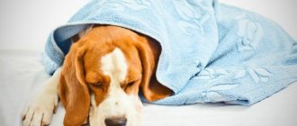
Dog after surgery
A dog’s normal recovery after surgery directly depends on how carefully the owner looks after it. The veterinarian will instruct the pet owner in detail how to care for him and what he needs to know, but there are also general rules for caring for a sick animal.
How is lymphoextravasate determined in dogs?
When diagnosing this disease, the doctor will take into account all the symptoms that we discussed above, carefully examining the animal. It is important to collect an anamnesis: has the dog been hit by a car or bicycle, been involved in a fight, or fallen from high surfaces ?
It is also necessary to exclude diseases that give similar symptoms:
- pathologies of the cardiovascular system
- kidney and urinary tract diseases
- swelling due to extremely severe exhaustion of the animal
- parasites, exposure to toxins can cause blood plasma to escape through damaged blood vessel walls
- extensive, systemic allergic reactions also cause increased vascular permeability.
To confirm the diagnosis, a sample of the liquid is taken with a special needle and its nature is determined. If it is lymph, then the liquid will be transparent, yellowish or reddish, slightly glowing in the light.
Postoperative nutrition for the animal
The attending veterinarian will provide specific recommendations on what the dog can do after surgery. This directly depends on the type of surgery and anesthesia.
The dog breeder wonders what to feed the dog after surgery? We offer the following:
- Feeding should be done little by little so as not to burden the body, since a lot of energy is spent on digesting food.
- After surgery, the dog does not eat or drink for several hours. This is especially true for operations performed on the peritoneum.
- There is no need to worry about your dog not going to the toilet after surgery. This is natural, because she doesn’t eat anything. And to avoid constipation, you need to stick to a diet. It is better to give dietary food, which is sold in special canned food. Hard food softens in warm water. This type of diet is observed for about 30 days. It is better to return to your normal eating rhythm gradually. To do this, the usual food is gradually mixed into the diet.
- In the postoperative period, it is preferable to give the dog broth, cottage cheese, kefir and porridge.
- There must be fresh drinking water near the dog.
- The owner must inform the treating veterinarian about negative reactions to food in the form of vomiting, diarrhea, constipation.
Diagnostic procedures
When diagnosing, all the same signs that we described above are taken into account. First, the veterinarian needs to look for marks on the dog's skin that indicate impacts. The owner is asked whether the dog was hit by a bicycle/car, whether it fought with other animals, whether it fell from obstacles during training...
Secondly, measures are being taken to identify other diseases, which, in some cases, can give similar symptoms:
- All diseases of the cardiovascular system.
- Pathologies of the urinary system.
- Extreme degree of exhaustion, in which hunger and cachectic edema are possible.
- Parasitic diseases, poisoning, in which the liquid part of the blood can escape through the walls of blood vessels.
- Generalized forms of allergic reactions. Histamine, abundantly released into the blood, also increases the porosity of blood vessels, the walls of which begin to resemble a colander.
In doubtful cases, the specialist simply takes an aspiration needle, through which a sample of fluid is taken from the swelling (suspected to be of a “lymphatic” nature). If there really was lymph in the cavity, then a clear, reddish or yellowish liquid is sucked into the syringe, which becomes slightly opalescent (shine) in the light.
If extravasation has formed on your dog’s ear, removing it is not particularly difficult for an experienced specialist. The dog is fixed in a special machine (general anesthesia is not required), the surface of the swelling is cut, and the liquid flowing from the cavity is pumped out with a syringe. The wound is washed with any standard antiseptic solution, after which the incision is “tacked” with several sutures. That's it, nothing else special is required.
The procedure is somewhat more complicated when internal, deep extravasations are treated. In mild cases, when the swelling is small and does not cause any particular concern due to its size (and also does not show a tendency to increase), one limits oneself to regular pumping out of liquid using an aspiration needle, and the surface of the swelling is occasionally lubricated with an alcohol tincture of iodine.
Sometimes the prescription of diuretics proves to be quite an effective method. Anti-inflammatory drugs, as a rule, do not need to be prescribed. The exception is those cases when lymph extravasation is caused by a strong blow, on the surface of which a deep abrasion, scratch, etc. remains. They are treated in the usual manner, preventing the contamination of the affected areas with pyogenic microflora.
Unfortunately, conservative treatment is not effective in all cases. If within several days there is no noticeable reduction in the “lymphatic” tumor, as well as in cases where it tends to increase, it is necessary to resort to a more radical operation.
In this case, the dog is immersed in anesthesia, the surface of the tumor is cut, and the liquid is removed with a syringe with an aspiration needle. If the incision had to be made long, and the extravasation itself is located deep in the tissues, it makes sense to leave a drainage impregnated with antiseptic compounds in the cavity. This is done in order to prevent the development of purulent inflammation. If necessary, the dog is prescribed antimicrobial drugs. Here's how to treat lymphoextravasate in dogs.
Important note! The main thing is never, under any circumstances, apply cold to the surface of the extravasate! This may cause the process to worsen. In addition, the sick animal must be in a state of absolute rest. If you take your dog for a walk, where he will actively run and frolic, or, worse, for training, the volume of the cavity will increase sharply, since lymph will be released several times faster during physical activity.
General information
A hematoma in this case is an accumulation of blood and ichor between the skin of the auricle and the cartilage that serves as the basis of the latter. The root cause of its occurrence is the rupture of a blood vessel. The larger it is, the more blood may end up in the hematoma cavity, and the greater the pain the dog will experience.
Note that this neoplasm appears (maximum) within an hour after the injury leading to damage to the blood vessel. At the same time, the ear swells, swells, becomes very painful, and the local temperature in this area increases noticeably. If nothing is done, the pain will subside within a few days, but the size of the ear will constantly increase due to the formation of scar tissue. As a result, the auricle will be deformed and disfigured.
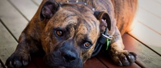
Are there any predisposed animals or breeds? In principle, any dog can suffer from ear hematoma, regardless of its gender and age, but in practice it turns out that most often this pathology occurs in animals with long, “droopy” ears (beagles, dachshunds, some Labradors), as well as , as we have already noted, dogs of fighting breeds, whose ears for some reason were not cropped.
All chronic ear infections, ear mites or allergies that cause the ears to become very itchy are most often the root cause of ear bruises. Animals will scratch their ears or constantly shake their head if they have ear mites. Please note that those dogs whose characteristics require the need to care for their ears (long-eared, long-haired breeds) are at increased risk. This is largely due to the owner’s failure to comply with the basic rules of asepsis and antiseptics.
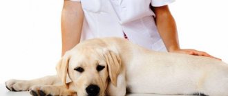
Diagnosis of the disease
If a dog has an ear hematoma, it is recommended to show the animal to a veterinarian. The specialist conducts an initial examination by palpation. As a rule, the dog reacts to such an examination quite aggressively: touching the affected area causes it discomfort and pain.
The doctor will also usually examine the animal's ear using an otoscope and take a swab from the pinna. The test is needed to determine the nature of the pathology: perhaps the otohematoma is caused by an infection or is of an allergic nature. In some cases, a puncture is performed.
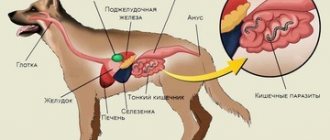
In addition, the veterinarian must find out whether the animal has any concomitant pathologies or diseases that could cause the otohematoma. For example, a hematoma-aneurysm can occur with lesions in the dog’s liver. In order for the specialist to get a complete impression of the nature of the pathology, you should tell him about all the symptoms accompanying the hematoma.
After examination and identification of the causes, the animal is prescribed appropriate treatment - we’ll talk about this later.
What to do?
Direct treatment of otohematoma in dogs can be carried out using several methods. Therapy will depend on the timeliness of its detection (the sooner the better), the size of the hematoma and the personal preferences of the veterinarian.
In most cases, simple surgery is required, which involves cutting the skin on the underside of the ear, inserting a drain to drain the accumulated blood, and then suturing it as often as possible. Typically, a bandage is placed on the auricle to protect the postoperative wound from contamination. During the same period, those diseases that led to organ injury must be identified and eliminated. Many owners are distrustful of the need for surgery, but only this method can guarantee that the auricle will not remain deformed. In addition, in this case the hematoma does not reappear (as a rule).
Disadvantages include the fact that short-haired dogs have noticeable scars on their ears, which does not suit all owners. This is especially true in cases where the dog has breeding value and constantly participates in exhibitions.
For such cases, there is another version of the operation, when the hematoma cavity is also opened, blood is also removed from it and drainage is inserted, but no stitches are applied. However, the veterinarian can still make a couple of stitches to secure the drainage in the open cavity. Accordingly, there are no scars left after such an intervention, and the auricle is not deformed.
This procedure requires more intensive care for the recovering dog, frequent replacement of bandages, and strict adherence to cleanliness. Since the wound is open, pathogenic microflora can easily enter it with all the ensuing consequences. Because of this, it is highly recommended that you take your dog to the veterinarian for bandages rather than doing it yourself.
(c) Veterinary center for the treatment and rehabilitation of animals “Zoostatus”.
Varshavskoe highway, 125 building 1.
Otohematoma
or pinna hematoma is a blood “pouch” that forms on the outside of the pinna, between the skin and cartilage in your pet’s pinna. As a rule, otohematoma is caused by damage to the ear due to scratching or occurs as a result of too intense and frequent shaking of the head, which leads to injury. Both dogs and cats suffer from ear hematomas, although dogs (especially those breeds that are prone to allergies and ear infections) are more susceptible to the disease.
Prevention
An owner who wants his four-legged friend not to be treated for such an unpleasant phenomenon as a hematoma must carefully monitor his pet. Regular examination of your pet’s ears and washing them, as well as accurate knowledge of allergens that are dangerous to your pet and the causes of otitis in dogs will help to avoid its occurrence. In addition, you need to avoid getting injured as a result of fighting and playing with other animals. It is important to understand that ignoring the appearance of a hematoma is dangerous for the life of your beloved pet.
Signs of hematoma
The presence of such damage can be determined by the following symptoms:
- Swelling of the damaged ear, the formation of a soft hemisphere filled with liquid.
- Increase in pet's body temperature.
- Pain at the location of the hematoma.
- Anxiety. The animal will constantly tilt its head towards the damaged ear; its appetite may decrease and interest in active games may disappear.
The dog may also experience severe itching. In this case, the animal will constantly scratch its ear, which will lead to additional traumatization. To prevent this from happening, it is recommended to contact a veterinarian and follow all his instructions.
How to eliminate medium and large hematomas?
Large tumors cannot be dealt with using the methods presented above. They will only aggravate the pet’s condition and can provoke the formation of suppuration. If your dog has a large hematoma full of fluid in his ear, he may need to have the blood drained with a syringe or have surgery. This treatment is carried out in a clinic. It is started 3-4 days after the hematoma appears, when the flow of blood into the cavity has already been completed and a blood clot has formed in it.
Pumping out with a syringe
For extensive uncomplicated injuries, the doctor may do without surgery. He will eliminate the hematoma with a syringe. This manipulation is carried out like this:
- The dog is fixed so that during the procedure he does not jerk his head (this will lead to injury to the affected ear). Anesthesia is not used for this treatment.
- The skin around the hematoma is treated with antiseptics.
- A puncture is made in the upper edge of the ear, through which the blood is sucked out. They try to eliminate the liquid without any residue, but if a small amount remains, it will resolve later.
- The syringe is changed, leaving the game in the wound. A solution of novocaine and antibiotics is injected into the cavity. Their type, as well as the dosage, are selected depending on the condition of the pet, the presence of complications, and the threat of pus.
- The ear is bandaged. The pet is given a hard collar to prevent it from scratching the damaged area.
Pumping out fluid from the cavity can be carried out in several sessions. Usually there are 5-7 of them. This is due to the fact that after the first treatment, blood soon begins to collect in the wound again. If the hematoma does not go away after such manipulation, surgical treatment is prescribed instead.
Carrying out the operation
Surgical intervention is prescribed in the most severe cases, when conservative methods do not give the desired effect. Do it like this:
- The dog is given general anesthesia. This is necessary so that the animal does not move its head during the operation.
- The affected ear is prepared for opening. The fur is shaved off and treated with chlorhexidine or another antiseptic. This is necessary to prevent infection from entering the wound.
- An incision is made along the ear. Remove accumulated liquid. If necessary, secure the damaged vessel with a ligature.
- The cavity is treated with an antiseptic.
- The ear is stitched. For this purpose, a rolled seam or through stitching of fabrics is used. The latter causes adhesive inflammation, allowing the cartilage and skin to grow together. If purulent processes are observed in the cavity, it is not sutured. The dog in the presented case may be left in the hospital for several days to ensure proper conditions of detention and regular treatment of damaged tissues. The wound will be stitched up only after pus stops accumulating in it. Partial suturing of the ear is performed if the vessel cannot be completely restored and the risk of re-hemorrhage remains.
After carrying out the presented manipulation, a collar is put on the dog so that it does not injure the damaged area of the skin. The animal is prescribed bandages, treatment of the ear with special antiseptic ointments and means to avoid scarring. In the future, the dog breeder will need to treat the dog at home.
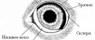
Ear structure
To understand how difficult it is to diagnose otohematoma, you should first understand the structure of the organ, which consists of the following components:
- outer ear;
- middle ear;
- inner ear;
- bone labyrinth;
- membranous labyrinth.
The structure of a dog's ear.
The outer part consists of the concha, the motor mechanism and the external auditory canal. The middle part includes the tympanic membrane, the tympanic membrane cavity, the chain of auditory ossicles, and the auditory tube. In the internal cavity there are receptors that are responsible for the level of hearing.
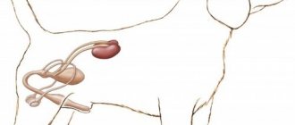
Bone labyrinth
The bony labyrinth is a system of hollow compartments that are located on the temporal part of the skull and serve to accommodate membranes.
- As for the membranous labyrinth, it is a whole system of adjacent hollow compartments consisting of connecting membranes. These compartments are filled with endolymph and in structure resemble an oval bladder containing three semicircular canals and the membranous canal.
- And also the named bladder serves as a container for the endolymphatic duct.
- The walls of the bladder consist of villous epithelium.
The bony labyrinth in a dog's ear contains membranes.