Types of liver cancer in dogs
The term neoplasia is used to refer to the process of uncontrolled cell growth. This name is derived from the words “neo” - new and “plasia” - development. The result is a tumor or neoplasm.
Each tumor can be:
- benign;
- malignant.
The first has clear boundaries, grows slowly and does not form metastases. And the second is often unpredictable, growing in any direction and penetrating into other tissues. All malignant neoplasms or tumors are considered cancer.
Did you know? According to statistics, 1 in 4 dogs will develop neoplasia at some stage in their life. Half of them in the age group over 10 years are diagnosed with cancer.
Liver cancer can occur at any time during your dog's life. It does not vary by breed or age group, although it tends to be more common in older animals. What makes the disease insidious is that its symptoms do not become particularly obvious. But in order to recognize them, you need to understand exactly how an organ like the liver works.
In humans, dogs and most creatures, the liver serves as a filter that helps the body eliminate toxins and potentially harmful substances. It is supplied with blood from the hepatic artery and portal vein, so it can perform its function as efficiently as possible. But for the same reason, the organ becomes more vulnerable, because cancer cells can enter it from another tissue using the bloodstream.
There are two types of liver cancer in dogs:
- primary - this means that the cancer process began in the liver itself;
- metastatic, when malignant cells have spread from another organ.
Metastatic is more common. It causes multiple tumors in the liver. But they are very likely to be benign. Primary cancer results from hepatocellular carcinoma (HCC).
There are several other types of cancer, including:
- bile duct cancer;
- neuroendocrine tumor;
- mesenchymal tumor (sarcoma).
Important! Any liver cancer can be accompanied by hemaniosarcoma (a tumor on the wall of a vessel). If it ruptures, it is always fatal. Then the pet dies almost instantly from the onset of bleeding.
Hepatocellular carcinoma can present in three different ways:
- massive - consists of one large tumor;
- nodular - represented by several neoplasms in different places of the liver;
- diffuse - covers the entire liver.
The most common form is the massive form. This type has a low frequency of metastasis and is very easy to remove, easier than diffuse or nodular. It must be remembered that without treatment, any form will begin to metastasize to the lymph nodes, lungs and other organs.
Metastatic (secondary) cancer means that the disease has spread from another organ.
It is usually associated with:
- pancreatic cancer;
- lymphoma;
- bowel cancer;
- thyroid cancer;
- fibrosarcoma;
- osteosarcoma;
- mast cell tumors;
- hemangiosarcoma;
- breast cancer;
- pheochromocytoma;
- transitional cell sarcoma.
Liver functions
The physiological role of the liver in the body of cattle is very important and multifaceted. The liver is the main organ involved in metabolism. Basic functions of the liver.
Function of bile formation and bile excretion. The formation and secretion of bile is one of the most important functions of the liver. Bile formation occurs as a result of the secretory activity of liver cells. The components of bile are bile pigments and acids, secretory products and excreta. Bile is secreted continuously, and bile secretion increases with increased blood supply to the liver.
Bile in the animal’s body enhances the effect of lipase, pancreatic and intestinal juices; emulsifies fats; helps neutralize acidic contents entering the intestines from the stomach, stimulates intestinal motility.
Neutralizing function. The liver is the main place for neutralizing substances of internal and external origin that are toxic to the body. Substances that are toxic to the body, such as cresol, skatole, indole, phenol, which are formed in the large intestine as a result of the decay of proteins, react in the liver with sulfuric and glucuronic acids and are converted into relatively harmless ester-sulfuric acids. Violation of the barrier function of the liver in cows leads to the development of nutritional toxemia.
Exchange function. Carbohydrate metabolism. The liver is the central organ in which most of the chemical processes associated with carbohydrate metabolism take place. Carbohydrates entering the intestines are converted into monosaccharides under the action of diabetic enzymes. Monosaccharides travel through the portal vein primarily to the liver. The liver retains a significant part of the sugar coming from the intestines and converts it into a special form of colloidal polysaccharide - glycogen, which is deposited in liver cells and serves as a reserve of carbohydrates in the body. Glycogen in the liver is formed with the participation of the hormone insulin; Without the influence of insulin, sugar is not able to pass into its active form and turn into glycogen.
The liver of healthy cows contains 3.72-3.9% glycogen. Cows with dystrophic processes in the liver have reduced glycogen reserves - 0.54-2.7%.
As a result of depletion of glycogen reserves, the urea-forming function of the liver is impaired. In addition, glucose is synthesized from body fat stores, which leads to the formation of excess ketone bodies, causing fatty liver degeneration.
In ruminants, carbohydrates are absorbed in the form of volatile fatty acids (acetic, propionic and butyric) - products of fiber fermentation. Highly productive cows have an increased need for glucose. This is explained by the rather high excretion of sugar in milk and its consumption by the mammary gland, heart and other organs.
Protein metabolism. The liver plays an important role in protein metabolism. Some plasma proteins are formed in it, in particular fibrinogen, prothrombin and others. Being a protein depot, the liver loses many times more protein during fasting than other organs. With liver damage, the total amount of proteins in the blood serum decreases.
Fat metabolism. In liver cells, saturated fatty acids are converted into unsaturated fatty acids. Phospholipoids are also formed there. The liver is actively involved in the breakdown of fats. The role of the liver in the synthesis of cholesterol has been established.
Pigment exchange. Bilirubin, both in physiological and pathological hemolysis, is formed from hemoglobin. The liver is not the only place where bilirubin is formed.
The liver is a depot for a number of vitamins. Vitamin A is synthesized in the liver from carotene.
The liver plays a big role in hematopoiesis, regulation of the metabolism of enzymes and vitamins, it is one of the regulators of the amount of blood circulating in the body, etc.
Why do cancerous tumors develop?
Most medical literature does not identify the underlying cause of liver cancer in dogs. But in some sources there is a theory that the absorption of pesticides and chemicals containing carcinogens can cause this disease.
Important! Most dogs do not show any signs of liver cancer until the tumor becomes too large and causes complex symptoms such as jaundice or weight loss.
There is also a theory that exposure to toxic substances disrupts the immune system and thus provokes the development of benign and malignant neoplasias. Toxins are found in dyes, household chemicals, and even some pet foods. Spoiled food and fungi can also be sources of these potentially harmful substances.
Jaundice in cattle.
Jaundice ( Jcterus ) is not an independent disease, but a syndrome characterized by yellow discoloration of visible mucous membranes, subcutaneous tissue and loose connective tissue.
With jaundice, in addition to bilirubin, other components of bile accumulate in the blood - bile acids, their salts, as well as cholesterol, which are toxic and cause the appearance of a number of signs in the animal (disorder of the functions of the nervous system, general weakness, rare pulse, itchy skin).
Pathologically, jaundice is divided into three forms: mechanical, parenchymal and hemolytic. It must be borne in mind that this division of jaundice is only a clinical scheme, since there are no permanent, unchanging boundaries between these forms. With various diseases, these boundaries are erased and one form invariably passes into another (G.V. Domrachev).
Mechanical (bilirubin) jaundice occurs in cattle when there are mechanical obstacles to the outflow of bile into the intestines (blockage of the bile ducts and small tubules by echinococcal blisters, neoplasms, parasites, cysts). Due to stagnation, bile accumulates in the excretory ducts of the lobules and stretches them. A pressure greater than the secretory pressure of the liver is created in the duct. As a result, after 15-20 minutes. bilirubin is found in the lymph, and somewhat later in the blood; The serum shows a rapid reaction to bilirubin after 24 hours. The accumulation of bilirubin in the blood carried through the liver in an animal is accompanied by severe jaundice and the appearance of it in large quantities in the urine, which also turns yellow. The foam that forms when shaking the urine will also have a yellow color. If bile does not enter the intestines, then it does not participate in the breakdown of fats. Hence, most of them are released with feces in the form of fatty acids and aldehydes. The feces of such an animal become gray-yellow with a strong odor, since they lack stercobilin, which delays putrefactive processes. The content of urobilin decreases with increasing mechanical obstruction, and with complete blockage it completely disappears from the urine. The course of obstructive jaundice depends on the cause that caused the blockage of the bile ducts. If there are stones in the bile ducts, partial obstruction occurs, which is eliminated after some time. Liver functions are not impaired in obstructive jaundice.
The development of parenchymal (bilirubine-urobilin) jaundice depends on damage to the liver cells - the liver parenchyma. Parenchymal jaundice accompanies infectious and septic diseases, feed and mineral poisoning. The liver with this jaundice is usually increased in volume. Bilirubin is secreted by liver cells into the bile ducts, into the lumen of the capillaries of the hepatic vein and is found in large quantities in the blood serum. During the study, bilirubin and urobilin are found in the urine, which indicates a decrease in the function of the liver to retain urobilin.
Hemolytic jaundice develops in animals due to increased formation of bilirubin during pathological hemolysis both in the bloodstream and in organs rich in reticuloendothelial elements. With hemolytic jaundice, there is an increase in stercobilin in the feces, which as a result have a dark yellow color. Hemolytic jaundice occurs in animals due to diseases in which the breakdown of red blood cells occurs, for example, under the influence of hemolytic poisons and various toxins, infectious equine anemia, leptospirosis, and large doses of arsenic entering the body.
How do symptoms appear?
Symptoms of liver cancer in dogs include:
- loss of appetite, which subsequently causes a sharp decrease in body weight;
- vomiting;
- light-colored feces;
- pain;
- bloated belly;
- cough, sometimes with blood;
- jaundice with accompanying symptoms - orange urine, pale gums, yellow irises of the eyes;
- extreme thirst;
- lethargy;
- high temperature.
All of the above symptoms, once they begin, will continue to progress.
Symptoms and manifestations
In most cases, in the early stages of development, liver pathologies are practically invisible. Possible liver diseases may be indicated by indirect signs:
- weakness;
- lethargy;
- poor appetite;
- thirst;
- weight loss.
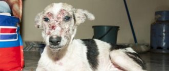
If extensive organ damage occurs, the symptoms become more pronounced. The dog may have:
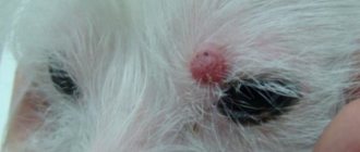
- jaundice, which is expressed by yellowing of the skin, mucous membranes, and whites of the eyes;
- nausea, vomiting;
- bowel dysfunction (constipation may alternate with diarrhea);
- change in the color of stool, it becomes light, almost colorless;
- urine is bright yellow, maybe brown, orange;
- flatulence;
- upon palpation in the right hypochondrium, an enlarged liver can be detected;
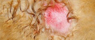
- peeling, itching of the skin;
- the appearance of pinpoint hemorrhages and increased temperature are possible;
- ascites is an accumulation of fluid in the peritoneum, accompanied by an increase in the volume of the abdomen.
In severe cases, there may be loss of vision and sense of smell. Coordination of movement is impaired, changes in behavior occur.
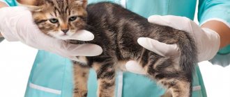
How is the diagnosis carried out?
First of all, a suspicion of a cancerous tumor arises from a veterinarian as a result of studying the dog’s medical history and general examination. Signs of jaundice or a swollen belly are noticeable immediately. And an enlarged liver can be detected by palpation.
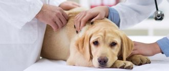
To confirm or refute his guess, the veterinarian will conduct a series of tests, such as:
- X-ray;
- blood tests;
- ultrasound examinations of internal organs.
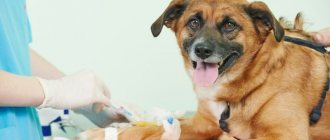
The oncologist also performs a liver biopsy. This is a detailed examination of tissue under a microscope. Unlike normal cells, cancer cells are characterized by an irregular shape, a darker nucleus and other abnormalities. This examination helps your doctor determine the type of cancer your dog has. And then discuss the treatment plan with the owner and select medications.
Forecast
After successful tumor removal, the long-term prognosis is good. The average life expectancy of the patient exceeds 4 years. Cats with operated bile duct adenoma also have a good prognosis.
Liver sarcoma has a poor prognosis. As a rule, during the initial visit to the clinic, distant metastases are detected (for example, in the lung tissue).
Thus, the prognosis depends on the type of tumor, the nature of the damage and the stage of the pathological process.
Read reviews about our veterinary center. Call the number and schedule a consultation right now or request a call back. (c) Veterinary center for the treatment and rehabilitation of animals “Zoostatus”. Varshavskoe highway, 125 building 1.
Treatment of liver cancer in dogs
Primary liver cancer in a dog seems like a dangerous diagnosis. But the liver is one of the few organs that is capable of regeneration. Therefore, removing part of it is not too difficult a problem for a massive form. Only diffuse, sometimes nodular forms and cancer that has metastasized to the liver from another organ will be problematic.
Important! Even if up to 75% of the liver is removed, the organ still continues to function and can recover.
Accordingly, treating the tumor by surgical removal is the most preferable option to solve the problem. And it will definitely be recommended for massive tumors in the liver. The remaining forms, unfortunately, have a poor prognosis. Presumably, the dog can live after confirmation of the diagnosis for 3–6 months. For tumors that cannot be removed, chemotherapy will be recommended. It will delay the progression of cancer.
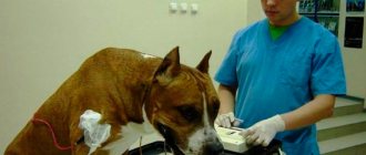
Carrying out the operation
As with humans, visualization of the tumor is necessary during the preparation for surgery.
For this use:
- X-ray;
- computed tomography;
- ultrasound;
- MRI.
Computed tomography is a more modern imaging option.
It allows you to obtain a three-dimensional model of the organ and the tumor in it. This is necessary in order to accurately perform the operation. Blood and urine tests will be required to get an idea of the dog's health and physical fitness, as well as any existing diseases. During the operation, the tumor and the space around it should be removed at a distance of 2 cm in any direction. If not everything can be removed, then additional chemotherapy or radiation will be used.
Chemotherapy and radiation
The goal of chemotherapy is to prevent metastasis or delay the appearance of metastases. Unlike people, dogs tolerate this type of treatment well and return to their normal lifestyle within 24 hours after it.
Did you know? Traditionally, chemotherapy is given intravenously, but there are drugs that are given by injection and even those that are taken orally.
Radiation therapy is used only for large tumors that cannot be removed. In this case, the part that is possible is surgically removed. And then using radiation to prevent or slow down the growth of cancer cells.
How to care for a sick dog
After surgery, your pet will be monitored in the intensive care unit. He will be prescribed medications to relieve the pain. Fluids and antibiotics are given through an intravenous line for several days after surgery to maintain hydration and prevent infection.
Nonsteroidal anti-inflammatory drugs (NSAIDs) are used to relieve pain. These medications also reduce inflammation, which results in pain.
Typical pain-relieving medications:
- "Metacam" at a dosage of 0.2 mg/kg subcutaneously;
- "Rimadyl" - 4 mg/kg;
- "Deramax" - 3–39 mg/kg;
- "Previcox" - 5 mg/kg.
These medications are suitable for reducing mild to moderate pain. Even if the dog is not in pain, these medications are still prescribed because they reduce inflammation.
Another class of therapeutic agents are drugs. They are prescribed for severe pain. The most common drug used these days is Tramadol.
And there are several reasons for this:
- It has an antidepressant effect.
- In moderate doses it is suitable for mild to moderate pain, and in large doses for severe pain.
But a high dose causes drowsiness.
To avoid this effect, Tramadol is combined with another anti-inflammatory drug. Another group of drugs is neurotransmitter modifier drugs. In this group:
- "Gabapentin" at a dosage of 10–60 mg/kg per day;
- "Amantadine" - 0.1 ml/kg;
- "Elavil" - 0.017–0.07 mg/kg.
They affect neurotransmitters, chemicals that cause the nerve to respond to pain signals. This group does not relieve inflammation, but it is effective in reducing pain. They are prescribed for long-term pain.
Did you know? It is believed that dogs can detect the scent of cancer cells. Therefore, they are used in medicine to detect lung, breast, skin, bladder and prostate cancer in people.
Daily blood tests are performed to look for signs of organ dysfunction and internal bleeding. If necessary, blood or plasma transfusions are performed. After discharge, painkillers and antibiotics will also be prescribed at home. Your pet will need an Elizabethan collar to prevent licking the incision after surgery. After 10–14 days, you need to show the dog to a veterinarian to monitor the postoperative condition.
It is very important for pets with liver cancer to follow a special diet. It must provide the body with substances that are lacking in an animal whose liver does not function properly.
In the product list:
- fish fat;
- turmeric;
- berries with a high content of vitamins C, E.
Cancer cells “feed” on carbohydrates. Therefore, there should be less of them in the diet. But they are indifferent to fat, so its share in the diet may increase. Talk to your veterinarian about the right diet for your pet. Also, tell him immediately if the dog is not eating or shows a significant decrease in appetite.
For dry food, Royal Canin Canine Hepatic or Hill's Prescription Diet l/d Liver Care is recommended for animals with liver disease. These are low protein foods. They are available in dry and wet form and are considered one of the best options for pets with liver disease.
Find out the symptoms and treatment of spleen cancer in a dog.
If you cook your own food, it should contain at least 50% meat. It can also be 50/50 potatoes with fish or 1/3 fish and 2/3 potatoes. You can add boiled carrots, pumpkin, beans, and eggs to the mixture. Break your meals into 4-5 small portions throughout the day rather than one large breakfast and dinner. This reduces the burden on the body when processing large pieces of food.
Treatment
If a single formation with a clear localization is detected, tumor removal is indicated.
Chemotherapy is carried out when a tumor is detected that is sensitive to the effects of drugs. To determine the type of tumor, a piece of the resulting tissue is sent for histological examination.
If an inoperable tumor is identified, a biopsy can be performed and further selection of a chemotherapy regimen that can slow down the progression of the pathology.
After surgery, the patient must remain in the hospital until all vital signs are completely stabilized. The animal will receive the required amount of painkillers, replacement infusion therapy, and antibiotic therapy. A low fat diet is indicated.
How long do dogs with liver cancer live?
How long a pet can live with cancer is probably the pet owner's top thought.
Life expectancy after liver cancer in dogs depends on several factors:
- Dogs with large tumors that have been removed have a survival rate 15 times higher than others. The average life expectancy for them after surgery is 3.8 years.
- For animals with a nodular or diffuse form, as well as with metastases, the average life expectancy is about 1 year. Such tumors cannot be completely removed and it all depends on how severe the cancer is in a particular pet.
Surgery and palliative care can prolong a pet's life, but it is also important for an owner to know the signs of late-stage liver cancer before death.
At this moment the following are observed:
- rapid weight loss;
- convulsions;
- loss of appetite;
- lethargy.
If you see these signs, you will need to make a decision about whether to keep your pet alive or let him go.
Diseases associated with medications
As noted earlier, liver diseases in animals can occur due to the use of certain potent drugs. Below are the most commonly diagnosed ones.
Cushing's disease
A neuroendocrine disease that occurs when there is an increased level of the stress hormone in the blood (cortisol). As a rule, the disease develops with prolonged and uncontrolled use of painkillers and hormonal drugs. Against the background of the emerging pathological process, hepatitis may develop.
Symptoms of Cushing's disease in dogs
Characteristic symptoms of the disease include:
- sagging abdomen;
- decreased muscle tone;
- general weakness of the body;
- frequent urination;
- increased thirst.
Once the diagnosis is confirmed, the veterinarian will prescribe medication, which is done in most cases. Drugs such as Cyproheptadine, Ketoconazole, Lysodren, Mitotane and others are prescribed. But if an adrenal tumor is detected, then the doctor is forced to resort to surgery, during which the affected adrenal gland is removed.
Dog with Cushing's disease
Ascites
Abdominal hydrops or ascites is a pathological condition in which free fluid accumulates in the abdominal cavity. In most cases, this disease is directly related to liver failure. Characteristic symptoms of ascites include an increase in the volume of the dog’s abdominal cavity: the animal looks like a large ball with legs and a tail. But this only happens in severe cases.
Basically, ascites causes increased gas formation, shortness of breath, tissue swelling, decreased activity, muscle atrophy and bouts of vomiting. If the pathology is accompanied by an infectious process, then antibacterial drugs are used in treatment. Also, for ascites, therapeutic abdominocentesis is performed, diuretics and oxygen therapy are prescribed. These measures will eliminate the unpleasant symptoms of the disease.
Ascites is a pathological condition in which free fluid accumulates in the abdominal cavity
Clinical significance
The authors currently believe that HNH itself causes little pain in affected dogs, but is of great value in the diagnostic evaluation of animals with HNH and other associated diseases. Macroscopic nodules can also be mistaken for neoplastic lesions, and the authors are aware of some cases in which it was recommended to euthanize animals on the basis of macroscopic infection on suspicion of metastatic neoplasia.
Dogs with HNH typically have elevated ALP concentrations, which often prolongs the period of laboratory testing and other diagnostic testing to determine the cause of persistently elevated serum ALP. Thus, HNH may distract the clinician from making a correct diagnosis in a dog with HNH symptoms and another condition. Hyperadrenocorticism is most common in study dogs with HNH and unexplained increases in ALP. The confusion is further complicated by the increasing frequency of ultrasound-guided needle biopsies, which are unable to show the characteristic nodular nature of the lesions, sending the attending physician in search of the causes of hepatic vacuolar pathological changes. Therefore, the main purpose of this article is to raise awareness among readers about HNH in older dogs.
Clinical diagnosis
HNH is a histologic diagnosis that requires liver tissue to demonstrate characteristic microscopic or macroscopic nodules. These nodules are composed of non-encapsulated hepatocytes, which can give a different morphological appearance, including relatively normal hepatocytes or with various vacuolar changes (fatty or hydropic degeneration). Vacuolar changes are thought to occur due to fatty infiltration, glycogen, and water accumulation (Meyer, 1996).
The latter indicates glycogen accumulation similar to the metabolic effect of glucocorticoids on hepatocytes in dogs. The nodules are characterized by a greater number of hepatocytes per unit area than in normal liver parenchyma, with a higher content of mitotic cells and a lower number of binucleated cells (Fabryetal., 1982). In severe cases, compression of the stromal structure can give a macroscopic and microscopic picture of cirrhosis, unless the liver is examined using a special stain. Fibrosis and inflammation are not components of nodular hyperplasia, but are present in cirrhosis. The surrounding parenchyma is often compressed by nodules, which changes the position of the sinusoidal portal tract and causes atrophy. Degenerative pathological changes such as focal hyperemia, vacuolation, hemosiderin deposits and lipofuscinosis may also be present in adjacent tissues (Johnson, 1995). Also often in connection with this is lipogranuloma (macrophages saturated with pigment), which in the presence of iron gives a bright color.
To make a diagnosis of HNH, the pathologist usually requires a wedge biopsy sample taken during surgery. Needle biopsy often does not provide sufficient liver tissue to demonstrate nodular hyperplasia and abnormalities in adjacent tissues. Core biopsy findings show only hepatocyte vacuolar changes, prompting the clinician to investigate a reactive or metabolic disorder such as hypercortisolism or lipidosis. Biopsy puncture testing also does not help distinguish HNH from hepatic neoplasia, in particular from hepatocellular carcinoma and adenoma.