- Classification of dislocations
- Symptoms of a dislocated paw in a dog
- Types of dislocations
- Treatment of dislocation
Dogs, especially young and healthy ones, are characterized by increased activity.
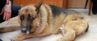
This behavior is completely incompatible with pain and injuries to the limbs. One of the most commonly diagnosed traumatic conditions is a dislocated dog's paw. A dislocation is a displacement of the bones of a particular joint without violating their integrity. Such an injury always entails damage to soft joint tissues - ligaments, tendons of adjacent muscles, blood vessels can tear, and the joint capsule is often seriously damaged. A dislocated joint entails dysfunction of the entire limb.
When to seek help from a veterinarian?
A dislocation in a dog is characterized by an abnormal location of the damaged organ or its individual part in relation to the physiological norm. The most common areas that are affected by dislocation are the kneecaps, hip joint, jaw and tail. In each case, the animal behaves differently and different symptoms are observed, indicating damage to one or another organ.
Signs of hip dislocation:
- tucking the paw under the body;
- swelling on the articular surface;
- change in joint shape;
- increasing howl;
- disobedience.
Symptoms of unilateral jaw dislocation:
- a slightly open or unnaturally “skewed” mouth;
- malocclusion;
- painful sensations in the mouth area;
- disobedience.
Signs of bilateral jaw dislocation:
- non-closing mouth;
- throwing the animal from side to side;
- quiet whining;
- aggressive behavior.
Symptoms of a luxated kneecap:
- swelling and swelling of the knee;
- the appearance of a “hanging” paw effect;
- the dog constantly lies in one place;
- severe pain in the knee area;
- aggression.
Signs of a dislocated tail:
- curvature of the tail;
- dislocation of the tail is characterized by its unnatural position;
- unpleasant sensations accompanied by quiet whining;
- weakness;
- manifestation of aggression towards others.
It is important to understand that when any of these signs are identified, you should carefully examine the animal and, if possible, provide first aid to alleviate the general condition, and then seek veterinary help.
Diseases of cats and dogs - diseases of the jaws and teeth (fracture, dislocation, periodontitis, caries, pulpitis)
Briefly: Diseases of the jaws and teeth (fracture, dislocation, periodontitis, caries, pulpitis) in cats and dogs are almost the same as in humans, and they are treated in approximately the same way. Read our article about how to treat your pets' teeth and jaws.
Home > Animals > Pets
Fracture of the lower jaw
Fractures of the chin, angle of the mandible and its alveolar process are differentiated. Fractures can be unilateral or bilateral, single, double, multiple, comminuted - without displacement and with displacement of fragments. Fractures passing through the dental edge are open and are often accompanied by damage to the teeth. Symphyseal fractures of the mandible (sagial) occur most easily.
Palpation and gentle squeezing of the lower jaw reveals pain in the fracture site, crepitus, and abnormal mobility. With symphyseal fractures, the dog retains its appetite, but cannot chew food. If the fractures are open, a wound and bleeding are detected. Fractures of the lower jaw can be complicated by its shortening and stiffness of the jaw joint.
Diagnosis is made based on symptoms and x-ray examination.
Immediately after a fracture occurs, it is necessary to stop the bleeding, prevent tongue retraction and asphyxia. Blood clots, foreign objects, and separated teeth are removed from the oral cavity. Therapeutic immobilization is achieved by using stainless steel wire splints that secure the teeth together. When twisting the wire, the ends must be turned inward. In the first week after treatment, solid foods are excluded from the diet and dairy products are given. If the fractures are open, the oral cavity is irrigated with a solution of furatsilin, and antibiotic or sulfonamide therapy is used. The wire splint is removed 30 days after surgery. It is recommended to take an x-ray first.
Dislocation of the lower jaw
It can occur when the dog’s mouth is forced to open very wide (grasping game), grasping large hard objects, falls, or bruises. It can be one- or two-sided. When dislocated, the articular head of the lower jaw jumps over the articular tubercle of the temporal bone and ends up in front of it. The integrity of the joint capsule is usually not compromised. The lower jaw is pushed forward and seems to be fixed, and drooling is observed. With a unilateral dislocation, the mouth is half-open, the lower jaw is skewed to the healthy side, and the dental bite changes dramatically.
Diagnosis is made based on symptoms and x-ray examination.
The lower jaw is adjusted. For this purpose, a stick is placed on the molars across the mouth, the jaw is grabbed with both hands so that the thumbs lie on the molars, and the rest cover it from the sides and below. By pressing on the teeth, the jaw is pulled down and moved from front to back - the head snaps into place with a characteristic click. After reduction, no solid food is given for 5-6 days.
Periodontitis
An inflammatory process that occurs in periodontal tissues located between the bone alveolus and the root of the tooth.
Tooth bruises, biting hard objects, bone cracks, transition of the inflammatory process from the gums or pulp. There are marginal periodontitis (periodontal damage along the edge of the dental alveoli), apical (periodontal damage in the area of the root apex) and diffuse (involves the entire root membrane). By its nature and course, periodontitis can be acute and chronic, aseptic and purulent.
Swelling of the gums around the tooth, pain on palpation and percussion, and tooth instability (diffuse destructive periodontitis) are detected. When the purulent process passes from the root membrane to the jaw, purulent osteomyelitis of the jaw occurs with the formation of fistulas.
The diagnosis is made based on symptoms and X-ray findings. Animals are fed soft food. The mouth is washed with a warm disinfectant solution (potassium permanganate 1:5000, 2% boric acid, furatsilin 1:5000). The gums around the tooth are lubricated with iodine glycerin. In case of diffuse septic periodontitis, the tooth is extracted. If periodontitis is chronic, sometimes there is an aggravation of the root sheath. Therefore, the removal of such a tooth may be accompanied by a jaw fracture.
Dental caries
It manifests itself as gradual destruction of the hard tissues of the tooth with the formation of a defect in the form of a depression (hollow). The process begins with a change in color, most often in a limited area, then the density of the dental tissue decreases here and a defect is formed. There are superficial, medium, deep (exposure of the dental pulp) and total caries (destruction of the entire crown). Caries can also begin from the pulp side.
The causes of caries are a violation of mineral metabolism (lack of calcium and phosphorus and, especially, such an important microelement for mineralization processes as fluoride), an acidic environment in the mouth (regurgitation during reflux of hydrochloric acid from the stomach, most often during sleep), and also a special microbial cenosis. Predisposing factors are tartar, tooth fractures, and insufficient hardness of the dental substance.
A frosty smell emanates from the mouth. There is a darkening of the tooth area (sometimes it is yellow), and the formation of a hollow. Caries is accompanied by periodic pain, drooling, difficulty chewing and lack of appetite. Formation of fistulas through the jaw outwards and osteomyelitis are possible.
The diagnosis is made taking into account the characteristic symptoms.
Foci of superficial caries are treated with a 1% solution of sodium fluoride, a solution of silver nitrate, and a fluoride-containing paste is rubbed into the tooth. Further progression of caries requires tooth extraction (after preliminary anesthesia). The dog is fixed on the operating table. The teeth of the upper jaw are anesthetized by injecting 2% novocaine into the infraorbital canal, lifting the upper lip with a finger. The infraorbital foramen is probed from the side of the mucous membrane of the vestibule of the mouth, slightly above the third premolar of the upper jaw. The needle is inserted along the lower edge of the recess, moving it parallel to the gum into the infraorbital canal to a depth of 2-3 cm. When positioned correctly, its tip penetrates to the sphenopalatine fossa.
To anesthetize the mandibular teeth, an anesthetic solution is injected into the mandibular foramen. The dog is fixed in a lying position with its mouth open, which is held in place with bandages. In the oral cavity, the ascending branch of the lower jaw is felt, 2 cm aboral to the posterior edge of the last molar, the mandibular foramen, which has the shape of a roller convex forward, is felt. The finger of the left hand is fixed at this point, and with the right hand an injection of anesthetic is made under the mucous membrane in the indicated place to a depth of 0.5 cm.
Before removing a tooth from the oral cavity, remove food residues that have accumulated between the teeth, rinse with a 1:500 solution of potassium permanganate, and lubricate the gums around the affected tooth with a 5% alcohol solution of iodine.
Using a scalpel, the gum is separated from the crown of the tooth. Using forceps, loosen the tooth using rotational movements. By pressing on the handles of the forceps, the tooth is removed. If there is pus in the alveolus, it is carefully removed and treated with an antiseptic solution.
Pulpitis
Inflammation of the dental pulp, located in its canal and including blood, lymphatic and nerve plexuses. The dental pulp is capable of responding to many pathological processes occurring in the animal’s body.
Inflammation of the dental pulp is often a consequence of its exposure after a fracture, caries, or premature wear of the crown. Sometimes the cause of pulpitis is the expansion of dentin tubules when it is exposed in a certain area. Microorganisms and their toxins penetrate into the dilated tubules, which leads to infection of the pulp. Prolonged exposure to a sufficiently strong irritant is accompanied by changes in the superficial and deep layers of the pulp, inflammatory hyperemia and leukocyte infiltration, which determine the nature of inflammation (acute and chronic, aseptic purulent, gangrenous and granulomatous pulpitis).
The lack of clear characteristic symptoms makes diagnosis difficult. Chewing of food on the side of the diseased tooth is impaired. Tapping a sore tooth causes a sharp painful reaction. With gangrenous pulpitis, a carious cavity and an exposed pulp of a dirty gray color are revealed, emitting a fetid odor. With granulomatous pulpitis, characterized by the proliferation of granulation tissue of the “wild meat” type, the process proceeds chronically, granulomatous growths protrude through the hole in the tooth.
In case of aseptic pulpitis, the animal is fed soft food, the circumference of the tooth neck is lubricated with iodine glycerin. A tooth affected by purulent, gangrenous or granulomatous pulpitis is removed.
(based on materials from “Diseases of Cats and Dogs”, edited by A.I. Mazurkevich, 1996)
How to help a dog with a dislocation?
If you discover an injury and the slightest suspicion of a dislocation of a particular joint, first of all, do not panic and calm down. Carefully examine your pet: make sure there are no tissue tears. Give him a drink of clean water - this will help him calm down and show less aggression towards you.
If your dog has injured its knee or hip joint, do everything possible to keep the animal from moving and lying on its side. For your own safety, first put on a muzzle (if it is not a dislocated jaw). In case of severe dislocation, the pet must be transported only on a well-stretched thick blanket or a rigid stretcher.
It is better to entrust this event to specialists and not touch the pet until they arrive. To ease the pain, bandage the injured finger or other part of the body with film, then with several layers of cloth and apply something cold (ideally ice).
Do not self-medicate under any circumstances, much less adjust the joint on your own. Call a veterinary surgeon to your home or take the animal to the nearest veterinary clinic.
How to identify a dislocation and provide emergency assistance
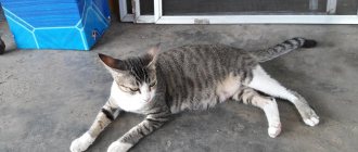
Naturally, the first symptom indicating problems with the musculoskeletal system is lameness or unnatural posture. A dislocation of the front paw is accompanied by a tightening of the limb and a “slumping” gait. If a dog cannot get up after sleeping or whines when lying down, there may be several possible diagnoses - dislocation of the hind paw, joint pathologies (arthritis), weakness, muscle strain. The main sign of a hip dislocation is that the paw is tucked in and “falls” inward; when injured or sprained, the dog also tucks the paw in, but keeps it in the correct axis.
So, what to do if your dog is injured and you suspect a dislocation:
- Do not panic - the injury is serious, painful, but not fatal.
- After providing first aid, be sure to take the dog to the clinic or call a veterinarian at home.
- Carefully examine the dog, make sure there are no tissue tears and the animal can move independently. If the hip joint is dislocated, do not let your pet walk! Calm and hold while lying on your side, the injured joint should be on top.
- Put a muzzle on your pet; even the most faithful and obedient dog can behave inappropriately when in severe pain.
- If necessary, the pet is carried on a mobile stretcher or a tightly stretched blanket.
- Do not try to reduce dislocations yourself! Closed injuries can be associated with internal bleeding and ligament ruptures.
- When fixing the joint with an elastic bandage, do not use a tight wrap. The elastic bandage tightens as you wear it. The ideal option is to apply a soft splint and fix the limb above and below the injured joint.
- Wrap the swollen joint in film and several layers of cloth, then apply ice - a basic measure that prevents bleeding and relieves pain.
- Monitor the temperature of the fixed limb; if the paw becomes colder, loosen the bandage.
- Do not demand an immediate answer from the veterinarian on how to treat your pet or a telephone consultation. Dislocation is confirmed by x-ray, with the exception of palpation in case of injury to the patella.
- If the dog whines or howls, it is necessary to give an anesthetic injection. In the absence of allergies and acute reactions, ketanol (ketanov), baralgin, rimadyl, or, in extreme cases, analgin, are suitable. If your dog has had acute reactions to medications and you don’t know what medications to give, use only veterinary medications, a universal pain reliever - Traumatine.
- Inject 1/2 dose according to the dog's weight, the pet should feel pain, otherwise it will begin to lean on the limb, which will worsen the condition.
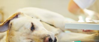
How are dislocations in dogs diagnosed?
At the clinic, an anamnesis is collected, the animal is examined, the damaged joint is palpated, and an X-ray examination is performed. Diagnostic measures can determine the presence or absence of a dislocation, as well as exclude or confirm a fracture in a finger, leg or tail.
The results of the x-ray will help determine the degree of violation of the integrity of the articular surface of the bones and prescribe appropriate treatment. In some cases, magnetic resonance or computed tomography may be necessary. This will allow you to more accurately see the clinical picture of the disease.
Classification of dislocations
- Congenital – pathology develops during intrauterine development. The born puppy is quite viable, but it is extremely rare to completely cure the disease.
- Traumatic - is a consequence of injuries that can occur when colliding with an obstacle at high speed, falling from a great height, or getting a limb stuck while moving. Often happens in car accidents.
- Pathological – arising as a complication of various pathologies of the musculoskeletal system, with thinning of cartilage or bone tissue.
- Paralytic - it is caused by atrophy of the muscle group supporting the joint.
- Habitual – this is the name for constantly occurring repeated dislocations, the cause of which is once stretched muscles or ligaments that weakly support the joint.
- Complicated - the bone is displaced, affecting nerve endings and important vessels.
- Unreducible - an old dislocation in which new tissues form between the articular heads.
How are dislocations in dogs treated?
The procedure for treating dislocations is carried out exclusively under general anesthesia. An anesthetic injection is given to completely relax the pet's muscles. The dislocation is then reduced to return the damaged ligament to its anatomically correct position. After which a special plaster bandage is applied to firmly fix the joint. The fixing bandage is left in place for up to 3 weeks, depending on the type and degree of dislocation.
In case of exacerbation and the appearance of temperature, chills, fever and other symptoms indicating the development of inflammatory processes, anti-inflammatory drugs are prescribed strictly according to the instructions of the treating veterinarian. For faster healing of ligaments and cartilage, chondroprotectors are prescribed.
After removing the fixing bandage, a course of physiotherapy is carried out, which lasts up to 5 weeks, depending on the speed of the animal’s rehabilitation. For quick recovery, regular walks in the forest, active swimming, physiotherapeutic warming and professional massage are recommended.
Prosthetics
The surgical method involves inserting an implant instead of the injured ligament. The method is most effective for medial luxation of the patella in a dog. Surgical treatment consists of anatomically correct fixation of the dislocated joint using a special surgical needle. Implant-supported prosthetics, as a rule, do not cause any particular inconvenience to its owner.
The pet is at complete rest with a surgical pin in the joint for 2-3 weeks. After complete healing, the wire is removed, and the resulting fibrous tissue provides support for the joint.
Diagnostic methods
To establish a diagnosis, a veterinary specialist only needs a clinical examination of the dog. The doctor carefully feels the furry patient’s limbs and conducts a test to determine mobility (flexes and straightens the limbs, listens for creaks and clicks when extending and bending the paws).
For a more accurate picture of the disease and the extent of the damage, you will need to perform an x-ray examination of the pet.
An x-ray will allow you to see whether there is a rupture of the round ligament or whether there are deep mechanical damage to the head of the femur.
Only after receiving the X-ray results does the doctor draw up a detailed treatment plan.