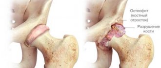
Signs of bone displacement
There may be several signs of musculoskeletal system disorders in the pelvic area. The owner needs to pay attention to the following deviations in the pet’s condition:
- inability to lean on the injured limb;
- immobility;
- lameness;
- painful sensations when touching the paw;
- incorrect anatomical location of the pelvic bones;
- swelling;
- heat in the damaged area;
- crunching, clicking in the paw when trying to bend or straighten it;
- the paw falls upward or is as if suspended;
- loss of appetite, apathy, weakness of the dog.
The limb may become a little shorter, longer, or its position is unnatural.
Patella luxation
This type of dislocation most often occurs in small and dwarf breeds of dogs, such as terriers and Spitz, poodles, as they have a genetic predisposition to such a dislocation.
Dislocation of the calyx as a result of injury occurs in larger breeds of dogs. Also, the displacement of the kneecap is often associated with age-related changes, since closer to old age the muscles of the body weaken and this type of injury is possible.
When a dog's kneecap dislocates, it can move both outward and inward - if the kneecap moves toward the abdomen, then this type is called a lateral dislocation, and in the opposite direction from the abdomen, it is a medial dislocation.
The last type - medial dislocation - is more common, small breeds of dogs are most susceptible to it, and for dwarf breeds it poses the greatest threat, as it can lead to irreversible consequences and the dog will lose mobility.
Classification
In order to diagnose a dislocation and determine treatment, it should be taken into account that doctors distinguish 4 degrees of severity of patella dislocation in dogs:
- The first degree is the easiest; after a knee injury, the cup will return to its place without consequences
- The second degree is characterized by an unnatural position when bending, and may fall into place in rare cases
- The third degree occurs during movement when bending and extending the paw; it requires surgical intervention, as it gets into place for a short time and loses its position again
- Fourth degree - with it the kneecap cannot return to its normal state and, accordingly, is always in a state of dislocation
Prolonged lack of treatment can lead to a chronic form of chromatia.
The symptoms of a luxated kneecap are as follows:
- The first stage is when the knee does not bend because the cup is not in its natural position, but it can be realigned. this condition rarely worsens and can be treated non-surgically
- At the second stage, the cartilage is severely damaged due to the sliding of the cup outside the joint, which leads to severe changes and the possible need for surgical intervention
- The third stage is a persistent dislocation, and without surgical intervention it is impossible to return the cup to its natural state, since even with successful reduction a relapse occurs and the cup flies out again
- At the fourth stage, the stability of the dislocation is also repeated, and even surgical intervention does not guarantee a 100% result
A luxated kneecap can be identified by the following external signs:
- the dog jumps or seems to shake off its paw during walks and exercise, but this does not bring it discomfort
- after a long rest, when the dog has not stood up for a long period, difficulty may be noticeable when trying to stand up; it is quite difficult to lean on its paws. After a long period of activity, nothing like this is observed.
- the dog moves its paw with less speed and range of motion
- the animal tries to constantly hold its paw in a bent state or press it to itself
- In the last severe stages, the joint becomes audible, since with any movement it crunches and clicks are heard
Diagnosis of a luxated patella occurs as follows:
It is necessary to take the pet to a veterinary clinic or invite a specialist to your home, after which the doctor will determine the position of the kneecap and determine whether it can be set without surgery.
Next, an x-ray must be taken in two positions: straight and lateral, which show the degree of dislocation and the presence of extraneous damage.
It is important to consider that exclusively medicinal treatment is based on the use of painkillers, and this is not always the right solution, since the dog stops experiencing pain and begins to actively use the damaged paw again. This is strictly contraindicated, as more damage occurs and the animal only harms itself.
Surgery for medial patellar luxation usually occurs in one of two versions:
The first method is plastic surgery, that is, removal of part of the cancellous bone, onto which fibrocartilage subsequently grows again.
The second is three-dimensional chondroplasty. This method is only suitable for pets up to six months old.
Since the first degree is not so dangerous, and when the kneecap is reset, the threat of re-dislocation is much less, you should be careful about the second and third degrees of dislocation.
With surgical intervention at these levels, a higher percentage of success and the absence of re-dislocation can be guaranteed. To do this, you should help your pet and carry out physical therapy with him, carefully monitoring the amount of exercise.
Consequences of injury, how dangerous it is
You cannot self-medicate . If examination and therapy are not carried out on time, this may result in the following consequences:
- contracture (tightening) of muscles;
- proliferation of cartilage tissue;
- complete destruction of the joint;
- disability of the animal;
- development of arthritis, polyarthritis with subsequent deformation of the pelvic bones.
With painful destruction of bone tissue, only open treatment will help. Its result will be complete immobilization or removal of damaged cartilage tissue.
Briefly about a ligament rupture on a dog’s hind leg
A cruciate ligament rupture in a dog of any breed is an injury to the knee joint, due to which the animal experiences severe pain and begins to limp on its hind leg. Most often, the ligament ruptures due to previously occurring pathological changes in the joint itself and the initially incorrect geometric location of the thigh and lower leg relative to each other. Very rarely, sudden movements during running and jumping become the main cause - usually this is a complementary factor.
Dog knee joint
In most cases, the anterior cruciate ligament (ACL) is torn, which causes instability and inflammation in the knee, and subsequently osteoarthritis, due to which the dog loses the ability to fully use its paw.
Osteoarthritis is an irreversible joint disease (degenerative change) that occurs when the cartilage tissue of the articular surface is damaged.
Click here to find out what diagnostics are performed for anterior cruciate ligament rupture.
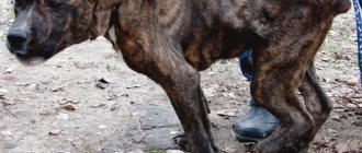
The dog is limping on its hind leg with no visible damage.
MAKE AN APPOINTMENT WITH THIS DOCTOR
What to do
If your dog whines for a long time and tucks its paw under its body, you should seek help from a veterinarian. What to do before then?
It is necessary to alleviate the pet’s condition: give painkillers, immobilize the injured limb, apply cold to the site of swelling.
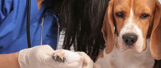
Survey
In a veterinary hospital, a dislocation is diagnosed during an external examination.
For a more accurate diagnosis, an x-ray examination is prescribed . This procedure will help the veterinarian make sure that the animal has a dislocation and not a fracture.
Conservative treatment
If the head has come out of the joint, it is possible to set it in a hospital setting under anesthesia. But such manipulation is rarely carried out: the probability of repeated loss is high, especially with dysplasia.
In dogs older than 5 years, the injury will certainly recur.
Reduction is carried out if the dog does not have concomitant diseases of the musculoskeletal system, skeletal system, in dogs under 5 years old, if the injury is not repeated.
It is important that no more than 24 hours have passed since the offset was received. Otherwise, muscle contracture occurs, cartilage tissue grows, and the animal will remain lame for life.
Operation
This treatment is considered the most effective method of restoring a damaged hip joint. There are practically no relapses after surgery.
There are several methods of open treatment for hip displacement:
- Ligament suturing and open joint reduction. After this, the damaged ligaments are shortened by suturing. The limb is immobilized, the animal is in complete rest for about a month. The main condition for recovery is proper rehabilitation in accordance with the doctor’s recommendations.
- Installation of a prosthesis: the damaged joint is replaced with a special implant. In this case, complete recovery occurs within a month, the animal feels well, and the foreign body does not cause any inconvenience.
- Arthroplasty is the restoration of a damaged joint. Conducted by a highly qualified doctor. Healing occurs quickly in a hospital setting. Within a week, complete restoration of the damaged tissue is observed.
During the postoperative period, the animal is prescribed painkillers and anti-inflammatory drugs. After surgery and rehabilitation, the probability of getting a new dislocation is no more than 1%.
Care during recovery
The complexity and duration of post-traumatic or postoperative treatment directly depends on the degree of complexity of the pathology and the specific technique that was used to restore the configuration and functions of the hip joint. However, in almost all cases, the animal is shown limited mobility (more or less strict), as well as fixation of the damaged part of the skeleton.
Important! Timely and reliable stabilization of the joint after its return to its original position is the main condition for ensuring a high treatment result and reducing the likelihood of re-dislocation.
Typically, the immediate rehabilitation period can last from 2 to 4 weeks. If the surgical intervention was accompanied by the use of metal knitting needles, pins and other temporary fixing structures, they are removed after the time stipulated for each specific treatment method.
Subsequently, the dog's motor activity should be restored gradually. Until further notice from the veterinarian, you can only walk with your pet on a leash in order to avoid new injuries and a possible relapse in such cases.
It is advisable that at first the animal undergoes a course of physiotherapy, mechanotherapy, manual therapy and special exercises (physical therapy). Swimming is especially recommended during the rehabilitation period, as well as running on sand or deep snow (depending on the time of year).
Dislocation: alarming symptoms
Damage to the hip joint in a dog can be congenital or acquired. A puppy may be injured at birth, but it cannot be determined until 3 months of age.
Older animals who are overweight and have a sedentary lifestyle are much more likely to suffer a dislocation than their counterparts who spend several hours a day outside.
As soon as the displacement is obtained, the dog begins to howl in pain and whine. The damaged area swells in a short time . When bending a limb, you can hear a characteristic crunch that occurs when bones rub.
The dog moves on 3 legs, the injured limb hangs down. Based on these signs, hip dislocation is preliminarily diagnosed at home without examination.
An accurate diagnosis is made at a veterinary hospital. In the first hours after injury, the injury is treated without surgery.
Diagnostic methods
To establish a diagnosis, a veterinary specialist only needs a clinical examination of the dog. The doctor carefully feels the furry patient’s limbs and conducts a test to determine mobility (flexes and straightens the limbs, listens for creaks and clicks when extending and bending the paws).
For a more accurate picture of the disease and the extent of the damage, you will need to perform an x-ray examination of the pet.
An x-ray will allow you to see whether there is a rupture of the round ligament or whether there are deep mechanical damage to the head of the femur.
Only after receiving the X-ray results does the doctor draw up a detailed treatment plan.
Is it possible to treat such an injury on your own?
It is impossible to treat a dislocation in a pet at home. This may cause the animal to become disabled. Only a doctor should be involved in realigning a damaged joint. The procedure takes place under general anesthesia in a hospital setting.
Non-surgical reduction of a dislocation is carried out when there is no ligament rupture, damage to the joint capsule, and the integrity of the muscles is not compromised.
Before a medical examination, the owner must do the following:
- Place the dog on the side opposite the injured area.
- Place a heating pad with ice on the injury site for about 15 minutes, no more. This will help relieve swelling.
- Try to immobilize the pet.
- Limit food, only drink.
If the dog is taken to the veterinary hospital within 2–3 hours, no pain medication is given. Otherwise, it is necessary to give an injection with an analgesic.
When is surgery needed?
After a full examination and x-ray, the veterinarian chooses treatment. In case of dislocations, surgery is prescribed.
This is done in the following cases:
- more than 24 hours have passed since the injury;
- the animal is more than 5 years old;
- the dog is overweight;
- medium and large breed dog;
- repeated damage;
- the joint is completely destroyed due to the disease;
- with dysplasia.
A serious injury that accompanies a dislocation of the hip joint is a ligament rupture. There is no conservative treatment in this case.
Treatment of hip dislocation
The sooner the dislocated joint is reduced, the better the long-term clinical result will be with a decrease in the likelihood of developing secondary arthrosis.
The optimal time for reduction is in the first hours after the injury.
If 12 to 24 hours have passed since the dislocation, the doctor may try to reduce the dislocation without surgery. Unfortunately, reduction of a dislocated hip joint using the closed reposition method most often does not bring a positive result; as a rule, a relapse occurs, and the femoral head comes out of the acetabulum again.
After reposition, the joint needs immobilization, which is difficult to achieve in an animal due to its anatomical structure and the inability to provide adequate rest.
The most reliable and effective treatment choice is surgery.
Surgical methods for treating dislocation
There are several methods that make it possible to ensure a complete recovery for the animal with a high degree of probability. The specialist chooses which method to perform reposition and fixation based on several factors, such as: the animal’s age, breed, body weight, chronic diseases, the nature and severity of the dislocation, and associated injuries.
Method of suturing the joint capsule (capsulorrhaphy)
The joint capsule is the main anatomical structure stabilizing the hip joint. When dislocations occur, a tear or rupture of the elements of the joint capsule occurs. After open reduction of the hip joint, the joint capsule is tightly sutured, and this serves as a stabilizing element (Fig. 1). After this operation, the animal must rest for 14 days.
Round ligament replacement method
The method consists of replacing the round ligament with a biocompatible implant (Fig. 2). This technique is similar to transarticular stabilization, but instead of a wire, a graft is inserted into the anatomical position of the round ligament. The remains of the round ligament are removed. The capsule is sutured and movements are limited for 7-10 days.
Transarticular stabilization method
The essence of this technique is to fix the hip joint in its correct anatomical position using a metal pin passed through the joint and fixing it to the bottom of the acetabulum (photo 2). During the postoperative period, the animal is limited in movement (short-term walks on a short leash, keeping in a limited space). After 2-3 weeks, the wire is removed, and the joint is stabilized due to the periarticular fibrous tissue formed during this period, and the functions of the limb are gradually restored.
Resection arthroplasty method
It consists of removing the head of the femur, after which the joint is stabilized due to the formation of fibrous tissue in the joint cavity (photo 3). It can be difficult for owners to understand how it is possible to restore the functions of a limb by removing a joint. In order to understand this, let’s pay attention to how the attachment of the thoracic limb to the skeleton is ensured; there is no joint between the scapula and the axial part of the skeleton and the connection is carried out exclusively through soft tissues (synsarcosis), the same thing happens with the pelvic limb after surgery. This surgical intervention is indicated for patients with dislocations due to hip dysplasia and a body weight of up to 15 kg. The undoubted advantage of this operation is the absence of any implants inside the joint. The recovery time depends mainly on the animal’s body weight; the smaller it is, the faster the restoration of limb functions occurs.
During the rehabilitation period in animals with dislocations of the hip joints, physiotherapy and swimming are recommended.
Caring for the animal after surgery
For 1-2 months after treatment, the pet is completely at rest. He is not given any physical activity.
After the operation, the animal is provided with comfortable conditions: a warm room, no obstacles to food and drink, limited space so that the dog’s movements are minimal.
Painkillers during this period
After surgery, the pet is regularly administered analgesics for the first weeks. This makes the animal feel better. The dog should sleep more and move less, which removes the load from the damaged area.
Causes of Anterior Cruciate Ligament Tear in Dogs
A complete rupture of the ACL can occur for three reasons:
- Knee injury due to excessive load
- Previously occurring anterior cruciate ligament tear
- Degenerative changes in the knee joint
Traumatic rupture of the anterior cruciate ligament
This is an acute injury that occurs suddenly during extreme physical activity. Damage occurs, as a rule, only in one paw (unilateral) due to the influence of the kinetic force of the body and movement.
The simplest example of a traumatic rupture of the anterior cruciate ligament is when the paw gets caught in some kind of depression while moving at high speed. That is, the dog falls into a hole, hole or snow at full speed, but does not stop, but continues to move forward by inertia. It turns out that the shin gets stuck in this hole for a moment, and the thigh continues to move - hyperextension of the knee joint occurs (hyperextension). In such a situation, the ligament may not withstand such a sudden heavy load and tear.
Excessive knee extension - hyperextension
Traumatic rupture of the ACL occurs, as a rule, in highly active dogs. As a rule, these are hunting or sporting dogs. But injury can also occur in some purebred dogs, such as English bulldogs. They are a bit of a clubfoot, so they are at increased risk of tearing the ligament if the tibia rotates excessively inward (internal rotation) during active movement.
At the same time, the thigh rotates outward and the lower leg rotates inward—internal rotation.
Cruciate ligament injuries can also occur in car accidents. When the blow falls on the knee, the knee hyperextends or the shin turns strongly inward, and the ligament breaks.
Cruciate ligament tear
If the injury does not immediately lead to a rupture of the ligament, then it can lead to a tear (partial rupture), in which the dog also begins to limp, but less pronounced. Without timely assistance, the tear almost always turns into a complete rupture of the anterior cruciate ligament, which results in severe lameness and pain.
Why does the ligament not completely tear?
The anterior cruciate ligament consists of two bundles:
- Anterior internal part (craniomedial). This part is more loaded, because it is involved in flexion and extension, so it is always damaged first.
- The posterior outer part (caudolateral) is involved only in extension and, as a rule, is more often preserved intact.
Thus, one bundle of the ligament remains intact, while the other is damaged - a partial rupture occurs.
If there is a tear, it is necessary to reduce physical activity as much as possible. Since restoration of structures (regeneration) takes 6-8 weeks, for many it becomes extremely difficult to keep the dog in a cage or small enclosure all this time. This is where owners make a huge mistake - they do not contact a veterinarian for diagnosis and treatment, but instead give the dog anti-inflammatory or painkillers on their own. The dog stops experiencing pain, begins to run and jump again, thereby completely tearing the damaged ligament.
If the dog is temporarily kept in strict conditions, the result of a ligament tear will not be a complete rupture, but restoration (regeneration) of the anterior cruciate ligament. This will save the animal from pain and lameness, and you from unplanned trips to the veterinary clinic and large expenses for surgical intervention.
Non-traumatic anterior cruciate ligament injuries
Most often, limping dogs are brought to the clinic not after car accidents or another hunt. As a rule, the owner says that the dog was simply playing in the yard or ran after the cat, and then yelped sharply and began limping.
A healthy cruciate ligament cannot be injured by normal acceleration. Those situations that owners describe as trauma are actually the last straw, which leads to the rupture of an already damaged ligament.
Rupture of the anterior cruciate ligament in dogs in 99% of cases is not due to injury at all, but due to chronic degenerative changes that occur after overuse or abnormal load on this ligament
In more than 60% of dogs, the ACL injury is bilateral, i.e. both right and left. But this does not mean that the ligaments are injured on both limbs at the same time, however, degenerative changes will occur in both.
Degenerative changes in the knee can occur for the following reasons:
- the angle of the tibial plateau in relation to the femur is too large - this is a very important factor;
- breed (anatomical) features of the joint structure (straightened knee, clubfoot, congenital defects, etc.).
- inflammation and destruction of knee joint structures;
- excess weight, since the knee joint of each dog is initially designed for a certain weight;
- constant heavy load (sports, hyperextension, etc.).
About the plateau angle, or one of the main reasons why dogs often tear their ACL
Nature has decreed that, anatomically, the dog’s thigh and lower leg are always at a certain angle relative to each other. Each breed has its own angle. Thus, German Shepherds always walk on bent limbs, while other dogs move exclusively on straight legs (Shar Pei, Chow Chow, Akita Inu, Mastiffs, etc.).
But no matter how the dog walks, there is still a bend in the knee joint. Therefore, even when the pet is simply standing, this already implies a complex balance between the flexors and extensors in the knee joint.
Schematic comparison of a human and dog joint
In humans, the knee joint at rest is quite stable, and the tension in the muscles of the lower extremities is low, since the moderate load is distributed evenly. We can lean against the wall, relax our leg muscles, and even fall asleep in this position without falling down. Our muscles will strain just to balance.
A dog will never be able to fall asleep standing up - the knee joints will bend and the animal will fall. This suggests that your pet cannot stand with relaxed limbs - he always has slight muscle tension (quadriceps - knee extensor). Consequently, the anterior cruciate ligament is also constantly under tension, even in a standing dog.
Why is the plateau angle in the dog’s knee joint so insidious?
A plateau is a plane with a relatively flat, clearly limited surface. In our case, we will talk about the tibial plateau (shin).
The angle of the tibial plateau is a fundamental element of the biomechanics of the knee joint. This angle is why some dogs are more prone to ACL tears and others less so. It also depends on this angle why, after an ACL injury, some dogs benefit from traditional or conservative treatment methods, while others do not, and why relapses or osteoarthritis develop.
The main cause of rupture of the anterior cruciate ligament is when the angle of insertion of the knee joint into the thigh was initially anatomically and physiologically incorrect from birth
Under such circumstances, the joint would have been injured for a long time, but the lateral ligaments “held” it until the last. But, unfortunately, over time the ligaments weaken and tear.
According to various studies, the angle of inclination of the tibial plateau in dogs is normally 18-30° (on average 23°), in humans 5-7° (very small), in a wolf 12-13°. Therefore, the closer the dog is to the wolf, the smaller the angle of inclination, therefore, the fewer problems with the cruciate ligament.
Comparison of the tibial plateau of humans and dogs
In humans, the tibial plateau (plate of the lower leg) is horizontal, so the thigh simply rests on it, like on a table - there is no need to make any effort. Consequently, the load from the thigh to the lower leg is transmitted in a straight line, which means the cruciate ligaments are not loaded.
But in a dog, the tibial plateau is not horizontal at all - the average inclination is 23° or more. Since the surface is inclined, the thigh should automatically slide off it. However, this does not happen because the cruciate ligament holds the bone firmly in place.
Function of the anterior cruciate ligament
Normally, the cruciate ligament works from year to year, but under constant heavy loads it experiences chronic stress, so degenerative changes begin - the ligament can weaken, become damaged and eventually rupture. Ligaments are loaded during hyperextension or excessive internal rotation (this is described above in the article). That is, getting injured in cases where there is a tendency for the thigh to shift relative to the lower leg.
Dogs with very straight knees
You should know that the more the knee joint extends, the greater the slope of the tibial plateau, therefore, the anterior cruciate ligament is more stretched and strained. And again in simple words:
The more the tibia is straightened relative to the hip (dogs that walk on straight legs), the more the tibial plateau deviates - this increases the load and significantly increases the risk of injury to the anterior cruciate ligament
And a little about knee extensors. In order for the knee to remain straight, you need constant tension in the quadriceps - the quadriceps muscle constantly, as it were, “pulls” the lower leg onto the thigh. And the more it pulls, the greater the load on the cruciate ligament. The calf muscle also “pulls” the thigh onto the lower leg, which also increases the load on the cruciate ligament.
Dogs with severely bent knees
German Shepherd
In dogs with strongly bent knees, on the contrary, the risk of injury is reduced, since the anatomical location of the knee at 90° transfers the main load to the posterior cruciate ligament, significantly unloading the anterior one. That is, when the knee joint is bent, the angle of inclination of the plateau, on the contrary, moves forward from the horizontal plane, and the tendency to move the tibia forward disappears.
Results
The angular characteristics of the knee joint are very important. It even happens that in some dogs, after a traumatic rupture, the knee heals without any veterinary care (without immobilizing the joint), and the animal does not even limp in the future. But these are just certain biomechanical features, which, unfortunately, are very rare in dogs. Therefore, if your pet has any gait disturbances, do not waste time - be sure to seek diagnosis and timely help from a veterinarian.
MAKE AN APPOINTMENT FOR YOUR PET WITH A DOCTOR
Inflammation and destruction of joint structures, or what is cruciate ligament disease
In addition to the tilt of the tibial plateau, there is another problem - degenerative changes in the ligament itself. This is the most common cause of anterior cruciate ligament rupture in dogs between 5 and 7 years of age. Such changes can be caused due to excess weight, repeated injuries, due to age-related changes and other things.
A healthy ligament, even after a rupture, is a strong “rope”, but a ligament that has undergone degenerative changes looks completely different. Examination of the tissue under a microscope shows that even in the absence of its complete rupture, the structure of collagen fibers is already significantly different - it resembles a shapeless substance. In fact, these connections practically no longer exist. Of course, such loose fabric cannot withstand the increased load and eventually breaks.
Overweight
Earlier in the article, we already raised the topic of excess weight, but it should be added that there is also an indirect mechanism for the influence of adipose tissue on other structures. The trouble is that fat produces pro-inflammatory hormones, which cause inflammation of the joints, skin, ears, kidneys, liver, heart, and other organs and tissues.
In an overweight animal, the joints become completely inflamed. Pro-inflammatory hormones enter the intra-articular fluid, where all joint structures are located, including the cruciate ligaments. Nutrition is disrupted, the ligament becomes thinner, becomes loose and soon breaks at the first unsuccessful movement of the dog.
Forgotten Traumas
Old injuries (especially those not treated in time) can leave their mark on the joint in the form of deformations, which eventually gradually lead to inflammation (arthritis).
Joint inflammation
A common cause of chronic inflammation of the cruciate ligament is osteoarthritis in the knee joint. This is a disease in which a large number of inflammatory mediators accumulate in the intraarticular fluid. They contribute to the inflammatory process of all living tissues with which they come into contact, including the cruciate ligaments, which are located in the intra-articular fluid.
Infectious (immune-mediated) inflammatory arthropathy also leads to rupture of the anterior cruciate ligament.
Elderly age
Age-related changes also make themselves felt - over the years, any tissue “wears out”, regenerates poorly and is poorly nourished. The knee joint is no exception. Any changes will certainly lead to the fact that the ligamentous apparatus will be greatly weakened, and the risk of rupture of the anterior cruciate ligament will become very high.
With degenerative changes, as a rule, ligament rupture occurs gradually, and clinical signs increase over time. Thus, due to a partial tear of the ACL, the dog initially begins to limp. But eventually, with a slight jump or during active games, it completely ruptures, which is manifested by clear clinical manifestations.
Young dogs
Unfortunately, young dogs can also have degenerative changes in the anterior cruciate ligament. The cause is congenital deformities of the knee joint itself or other pathologies of the pelvic limb (for example, hip dysplasia or luxation of the patella). Due to improper stress on the ligament, it constantly undergoes changes and soon ruptures.
What are the types of dislocations?
Damage to the hip joint is divided into congenital and acquired.
Birth dislocation occurs in puppies as a result of improper development of bone tissue during the prenatal period. In this case, treatment is not carried out.
The dog suffers an acquired dislocation due to mechanical damage or disease. This species is found only in adults.
Acquired dislocation is divided into the following groups:
- unreducible (repeated injuries);
- paralytic (damage leading to atrophy of the muscles around the joint);
- complicated (with rupture of ligaments or muscles).
Traumatic dislocation can be complete or incomplete . With complete displacement, the joint capsule is completely ruptured, the bones do not touch each other, and the cartilage tissue is completely damaged.
Predisposing factors of pathology
What circumstances lead to hip dislocation in dogs? If any of the above structures are damaged, dislocation will not take long to occur. Most often, serious mechanical injuries lead to this outcome. In more rare cases, the immediate cause is dysplasia. Fortunately, both hips are extremely rarely affected (no more than 5-6% of cases with very serious injuries). But there are other predisposing factors that can lead to hip dislocation.
Dislocation in puppies
In puppies, such an injury is considered congenital. This indicates a change in the composition of bone tissue. This deviation cannot be treated.
As soon as the puppy is 2-3 months old, it must be taken to the veterinarian and consulted. After the x-ray, a decision will be made to replace the dog with prosthetics, but at an older age. Most often, such animals are euthanized.
If the puppy's injury was acquired, its treatment is similar to that of adult dogs. The puppy is growing quickly and prosthetic surgery will be delayed until he is older.
First aid
If your dog shows symptoms of a dislocation, you should not panic, since the injury, although serious, is not life-threatening if treated in a timely manner. The main thing is to provide first aid correctly and immediately call a veterinarian or take the animal to the clinic.
First of all, you need to put a muzzle on your pet, since any dog in pain shock can become aggressive and unpredictable. Then the algorithm of actions should be as follows:
- carefully lay the pet down with the damaged joint facing up and inspect for tissue tears;
- do not allow him to walk, but, if necessary, carry him on a stretcher or blanket;
- fix the joint with an elastic bandage, ideally with the application of a soft splint with fastening above and below the joint;
- wrap the injured area with film, then with cloth and apply cold.
You can’t straighten a dog’s dislocated paw yourself! Improper actions can cause significant harm and cause severe pain to the animal, especially if the injury is accompanied by ligament rupture and internal bleeding.
Before contacting a veterinarian, it is necessary to monitor the temperature of the limb with a fixing bandage. If she gets cold, then the bandage should be loosened.
In case of severe pain syndrome, in which the animal whines or howls, it is recommended to inject an anesthetic drug. Such traditional “human” analgesics as Ketanov, Baralgin or Analgin are suitable for this. If your pet is allergic to such medications, only zoo medications, in particular Travmatin, can be injected.
Rehabilitation
After surgery, the pet needs to fully or partially restore bone and cartilage tissue.
Supplements rich in calcium and collagen are introduced into the dog’s diet. X-rays are taken regularly to monitor the restoration of tissue around the joint.
After 6 months, the dog can increase physical activity, but this must be done gradually.
Disease prevention
The essence of preventive measures to prevent dysplasia in dogs depends on the stage at which they are required. It is necessary to think about the absence of a disease in a puppy before purchasing it. When choosing a large breed dog, you need to make sure that its parents have been tested for dysplasia and showed negative results (grade A). A certificate about this is provided by the breeder along with other documents. Although even this will not give a complete guarantee that the disease will not manifest itself in the future.
It is simply impossible to determine dysplasia in a puppy under the age of 6 months (and sometimes older). But if the dog has a predisposition, the disease will certainly manifest itself later. Therefore, further prevention consists of minimizing the risk of its occurrence or development of consequences. Preventive measures include a balanced diet and adequate exercise. With this approach, it is quite possible to stop the development of the disease, even if a pathological process has begun in the puppy’s joints.
If a large breed dog is overfed from childhood, which leads to rapid weight gain, and at the same time subjected to excessive training, then all this together significantly increases the load on sore joints and can cause irreparable harm to the animal. Any dog requires attention and care, especially if it is a large breed that is at risk for joint diseases. However, you need to know that dysplasia is not a death sentence. You can save your pet if you notice the problem in a timely manner and provide it with proper treatment.
You can also contact our site's staff veterinarian, who will respond to them as soon as possible in the comment box below.