Liver tumors in dogs and cats
Authors: Julius M. Liptak, BVSc, MVetClinStud, FACVSc*; William S. Dernell, DVM, MS, DACVS; Stephen J. Withrow, DVM, DACVS, DACVIM (oncology). Colorado State University.
Liver tumors can be primary or metastatic. Metastatic neoplasms of the hepatobiliary system are more common than primary ones. Primary liver tumors of dogs and cats are divided into 4 categories: hepatocellular, bile duct, mesenchymal and neuroendocrine. Malignant variants of these tumors are more common in dogs, while benign neoplasms, in particular bile duct cystadenoma, are more common in cats. Morphologically, primary liver tumors are classified as solid, nodular, or diffuse. Solid tumors have a better prognosis than nodular, diffuse, or metastatic tumors because surgical resection and cure are possible, especially for hepatocellular carcinoma in dogs and bile duct adenoma and myelolipoma in cats. Conversely, for nodular, diffuse and metastatic liver tumors, treatment options are limited, since surgical removal is not always possible and other forms of treatment have not been developed.
Primary liver tumors are not very common and account for 0.6 - 1.5% of all tumors in dogs and 1.0 - 2.9% in cats, but in cats they account for up to 6.9% of tumors not related to tumors of the hematopoietic system. 1–4 Metastatic disease, particularly metastases from splenic, pancreatic, and gastrointestinal cancers, is more common and is 2.5 times more common than primary tumors in dogs. 1,2 Liver involvement is also possible in other malignant processes, such as lymphoma, malignant histiocytosis, and systemic mastocytosis. 2.3
Primary malignant liver tumors in cats and dogs are divided into 4 main categories: hepatocellular tumors, bile duct tumors, neuroendocrine (or carcinoid) and mesenchymal tumors. 4
More details
Treatment
Surgical treatment and transplantation
If the patient meets the Milan criteria, surgical treatment is recommended by liver transplantation.
Liver resection is becoming the procedure of choice for patients with tumors smaller than 5 cm in the absence of cirrhosis. These patients can often tolerate resection of up to 50% of the total liver volume. Resection of more than two segments is contraindicated in patients with grade B or C cirrhosis. However, among patients who undergo successful resection, long-term survival is possible, with 5-year survival rates reaching 74% in patients without significant decompensation.
After liver resection, 75% of patients develop intrahepatic recurrence within 5 years.
Local ablative therapy abroad today is limited to radiofrequency ablation (RFA). Other methods, as practice has shown, are ineffective. The scope of application of RFA is tumors less than 4 cm in diameter and palliative care.
The method of chemoembolization (closure of the intrahepatic artery with gelatin foam), previously widely used in the treatment of small tumors, is now also increasingly used in palliative interventions.
Radiation therapy
Today in Europe, mainly two radiotherapy technologies are used for different types of HCC.
- Brachytherapy using TheraSphere technology uses glass beads ranging in size from 20 to 40 microns with radioactive yttrium, which are delivered to the tumor through the vascular bed. Radiation therapy is then carried out for 10-12 days with a total dose of about 150 Gy. The maximum permissible distance is 1 cm.
- Stereotactic computer-assisted radiosurgery using the CyberKnife technique is used as palliative treatment to reduce tumor size when surgical resection is not possible.
Targeted therapy
Sorafenib, a multitargeted oral kinase inhibitor, has been shown to prolong life in patients with hepatocellular carcinoma.
The agent, which targets multiple pathways including those encoded by VEGFR, PDGFR, KIT, FLT-3 and RET, was compared to placebo in a study of 602 patients. Median survival was significantly increased to 10.7 from 7.9 months, and time to progression was 5.5 months compared with 2.8 months in the placebo group.
Regorafenib (Stivarga) and cabozantinib (Cabometyx) are inhibitors of multiple tyrosine kinases, including RET, MET, and VEGFR-2. They showed good therapeutic activity in patients with poor response rates to sorafenib.
The mTOR inhibitor sirolimus, which may be useful in the treatment of both cholangiocellular carcinoma and hepatocellular carcinoma, is also being studied in clinical trials.
Immunotherapy
Immune checkpoint inhibitors have been shown to result in long-lasting tumor shrinkage, leading to significant overall survival benefits over standard therapy in some types of cancer. Blocking the PD-1/(PD-L1, PD-L2) signaling pathway has shown promising results in patients with hepatocellular carcinoma.
Nivolumab (Opdivo) has shown good results in the treatment of hepatocellular carcinoma, including in patients who have previously received sorafenib. The overall confirmed response rate was 14.3% (95% CI: 9.2, 20.8), with 3 complete responses and 19 partial responses. Duration of response ranged from 3.2 to 38.2+ months; 91% of respondents had responses lasting 6 months or more, and 55% had responses lasting 12 months or more.
Pembrolizumab (Keytruda) is another PD-1 inhibitor that is used abroad to treat HCC in patients previously treated with sorafenib.
Tumors of the pancreas and liver in cats.
Oncological diseases (malignant tumors) affecting the functions of the liver and/or pancreas are, fortunately, quite rare in cats.
Symptoms of malignant tumors may include jaundice (due to obstruction of bile flow), depression, weight loss, vomiting and bloating (due to the tumor itself or due to fluid accumulation in the abdominal cavity).
Most often, adenocarcinoma develops in the pancreas; other types of tumors are less common - adenoma, lymphosarcoma, squamous cell carcinoma, lymphangiosarcoma and spindle cell sarcoma. Diagnosing pancreatic tumors in cats is not easy, as symptoms can relate to many diseases, and blood tests may be normal. The most common change visible on ultrasound. is the appearance of nodules or dense formations of various sizes in or near the pancreas. Nodules in the liver can signal a variety of diseases, so cytology or histology of the biopsy material is necessary to correctly diagnose the nature of the tumors.
The prognosis for treatment of tumors of the pancreas and liver (bile ducts) is very unfavorable; such tumors respond poorly to existing treatment methods.
In addition to malignant ones, cats may also develop benign tumors of the liver or pancreas, which may not cause any significant clinical signs of the disease.
Site news
03/19/2016 How to prevent aggression towards other cats.
03/12/2016 Why are cats afraid of water?
More details
Liver tumor in animals
Liver tumors in animals are a rare group of tumors, and are recorded in no more than one percent of cases. Most animals arrive for examination at a veterinary clinic already with an advanced form of the disease.
Benign forms of tumors occur with little symptoms, and sometimes completely asymptomatic, and are formed both from epitolyal tissue (hepatocellular adenoma) and from vascular and stromal elements (hemangioma).
Hepatocellular adenoma is clinically asymptomatic, which develops from hepatocytes and is most often limited to the capsule. If the tumor grows strongly and vigorously, it may rupture with bleeding and severe damage to blood vessels.
Focal nodular hyperplasia of the liver is a benign liver tumor in animals, which is a dense scar-connective tissue in the central part, and at the edges it is represented by nodular-transformed hepatocellular tissue. Sometimes called “focal cirrhosis” - since, as a rule, it does not develop in a cirrhotic liver.
Regenerative nodular hyperplasia of the liver is very often combined with focal nodular hyperplasia of the liver, but unlike it, the regenerative form contains connective tissue elements in smaller quantities. As the tumor grows, large bile ducts, the gallbladder, and large branches of the portal vein are most often affected, and cirrhotic changes in the liver are not observed.
Liver hemangioma is an asymptomatic benign liver tumor in animals, which usually originates from the venous elements of the liver. It is one of the most common and recognizable benign tumors.
According to statistics, most of all registered primary malignant tumors are hepatocellular cancer, followed by tumors of the bile ducts, and in the remaining cases carcinoid and fibrosarcoma occur.
In cats, benign liver degeneration is most common.
More details
Diagnostics
Laboratory research
Alpha fetoprotein (AFP) is elevated in 75% of cases. The level of elevation is inversely correlated with prognosis. Elevations greater than 400 ng/mL predict hepatocellular carcinoma with a specificity greater than 95%. In the setting of tumor growth, cirrhosis, and the absence of acute hepatitis, many centers use levels greater than 1000 ng/mL as presumptive evidence of hepatocellular carcinoma (without biopsy).
AFP is not suitable for screening in active hepatitis due to the high rate of false positive results!
Desamma-carboxy-prothrombin (DCP) has been studied as a biomarker for the early diagnosis of hepatocellular carcinoma. The sensitivity and specificity of DCP during the diagnosis of hepatocellular carcinoma were found to be 74% and 86%, respectively, at a cutoff of 40 MAU/mL and 43% and 100%, respectively, at a cutoff of 150 MAU/mL. Twelve months before diagnosis, sensitivity and specificity at a cutoff of 40 MAU/ml were 43% and 94%.
The functional state of the liver and the degree of cirrhosis are determined by the following tests:
- total bilirubin;
- aspartate aminotransferase (AST);
- alkaline phosphatase;
- albumen;
- prothrombin time.
Medical Imaging Techniques
The simplest option is ultrasound. But its scope of use is limited. This method is powerless for small nodular and diffuse carcinomas. Moreover, it (in the Doppler version) can be successfully used to determine the degree of blockage of the portal vein.
Almost all the necessary diagnostic information can be obtained from an MRI scan. This method shows well the structure of liver tissue and allows you to detect small tumor formations at the earliest stages.
Biopsy
This method serves to definitively confirm the diagnosis of HCC and to determine its type.
Liver cancer in cats
Primary liver cancer is rare in cats, accounting for only 2% of all cancer cases. Much more often, cats suffer from secondary liver damage, in which malignant cells enter the liver from other organs, or through the lymphatic/circulatory systems.
Author: Bushow Karina
Breed does not play a role in the development of liver cancer in cats. The disease is most often observed in animals over 10 years of age.
Causes of liver cancer in cats:
contact with toxic substances.
Diagnostics
The veterinarian will conduct a physical examination of the animal, take a complete blood count, urine test, and count blood cells. X-rays are used to determine the location of a cancerous tumor. To make an accurate diagnosis, the doctor will do a biopsy - take a sample of liver tissue. Needle biopsy for liver cancer in cats is not recommended.
Treatment for liver cancer in cats depends on the type of tumor, its size and the presence of metastases in the body. The main treatment method is surgical removal of the affected part of the liver. A veterinarian can remove up to 75% of this organ without much harm to the cat's health, since the liver has a high ability to regenerate. If there is metastasis in the animal's body, chemotherapy may be needed in addition to surgery.
In the absence of metastases and the ability to completely remove the cancerous tumor, the animal in most cases returns to normal life. With a metastatic form of cancer, the life expectancy of an animal after treatment is about a year. The least favorable prognosis is for animals whose liver is so damaged that surgery is impossible.
Prevention
Avoid contact of your cat with pesticides, paints and any other toxic substances. Feed your pet only high-quality food without added chemicals. Bring your animal to the veterinarian regularly for preventive examinations.
More details
Etiology
The incidence of cancer among cats is increasing every year.
Experts attribute this to pets living near people who do not always take care of the animal in the right way. People feed their cats low-quality dry food, which contains a huge amount of preservatives and chemicals that can have a carcinogenic effect. Carcinogens affect the genome of the cell, and it begins to mutate, multiply rapidly and gradually turn into a full-fledged tumor.
According to another theory, the cause of cancer lies in its viral nature. Penetrating into a macroorganism, an oncovirus (the number of species is more than 100) remains in suspended animation, or its growth blocks the immune system. Any factor can serve as an impetus for the activation of cancer: stress, poor diet, frequent illnesses, infections and parasites. In such cases, the immune system fights the problem present, and at this moment the malignant cells begin to actively divide.
Cancer in cats
Cancer in cats is responsible for half of all cat deaths after 10 years of age. A malignant tumor in animals is capable of metastasizing, which quickly affects all healthy cells of the body. Cancer in cats can be detected in advance, in which case there is a likelihood of the animal recovering and increasing its lifespan.
Symptoms of cancer in a cat
If you notice the following symptoms in your cat, you should sound the alarm:
- tumors in the mouth and nasal cavity can be malignant. The appearance of a tumor is indicated by an unpleasant odor from the animal’s mouth, bleeding gums, difficulty breathing, difficulty swallowing;
- swollen lymph nodes may be a sign of lymphoma;
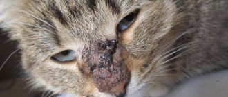
- pigment spots on the skin, non-healing ulcers can be a sign of skin cancer in cats;
- loss of weight, appetite, thirst;
- bleeding (of any organ);
- decreased activity, apathy;
- formations in the ears;
- education in the mammary glands.
Treatment
Treatment for cancer in cats will depend on the type of cancer, its degree, and the general condition of the animal. Chemotherapy, radiation, immunotherapy, and surgery may be prescribed. You need to understand that the drugs used to treat cancer are very active, and the animal will feel very bad after each procedure, but this does not mean that the treatment is harmful. The improvement will not be noticeable immediately. The cat may vomit, it may lie and sleep most of the time, and it may meow restlessly. The animal's behavior after the procedure should be discussed with a doctor, who will explain whether this is normal and whether it is worth interrupting treatment.
The doctor will also advise on proper nutrition for a cat with cancer. Dietary considerations will depend on what type of cancer the cat has. Many cats with liver cancer refuse to eat at all. In this case, it is advised to feed the cat with a syringe (without a needle, of course), with soft pureed food. You should not allow your cat to lose weight. The doctor may prescribe painkillers and substances to make food easier to digest, or may prescribe injections or even IVs.