What is tracheal collapse in dogs?
The trachea (literally the ancient Greek “τραχεῖα” is translated as a rough tube) is an organ related to the respiratory system and is responsible for ensuring the flow of air from the nasopharynx or throat directly into the lungs. In humans, dogs and all other vertebrates, the trachea is located between the bronchi and the larynx.
Externally, the trachea is a hollow tube formed by hyaline half-rings of cartilage tissue and consisting of two parts - cervical and thoracic. The thoracic part, or tracheal membrane, facing the spine, should close the cartilaginous rings that are not completely connected to each other. The membrane is based on connective tissue and smooth muscle. It is thanks to the large amount of cartilage and muscles that the trachea in its normal state is very flexible and elastic. In addition to moving air from the upper respiratory tract to the lower, it is also designed to retain on its surface the smallest particles of dirt, dust, pathogenic microflora and other foreign impurities present in the atmosphere, which are then expelled from the larynx to the outside through coughing.
If the cartilaginous rings for some reason (for example, due to chronic inflammation) lose their rigidity and elasticity, the trachea sags in this area. As a result of this deformation, there is a narrowing of the lumen that should be present in the tube. The tracheal membrane acquires unnatural mobility, sagging at the inlet and protruding during exhalation, which prevents the normal movement of air through the tube and, accordingly, makes breathing difficult. This condition is called tracheal prolapse (from the Latin “prolapsus” - prolapse).
At the moment when the progressive prolapse acquires life-threatening proportions for the animal (the dog begins to choke), a more serious stage of the disease occurs, called tracheal collapse (from the Latin “collapsus” - fallen).
Important! Prolapse and tracheal collapse are descriptions of the same pathological process, but in the first case we are talking about the anatomical characteristics of the problem, in the second, about the degree of its clinical manifestation. In other words, prolapse is a process, and collapse is the result of this process.
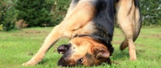
The flattening and flattening of the trachea during collapse always has a dorsoventral orientation, that is, directed from top to bottom, from the pharynx to the spine. The respiratory lumen in the tube, instead of being round, as it should be, becomes almost flat, its opposite walls begin to touch each other, as a result, suffocation occurs - at first weak, and then complete.
Medical science calls the narrowing of the lumen of any normally hollow organ stenosis (from the ancient Greek “στενός” - narrow) or stricture (from the Latin “strictura” - to shrink). Depending on the degree of development of stenosis, 4 stages of pathology are distinguished:
- first - closing the lumen by a quarter;
- the second - half;
- third - three quarters;
- fourth - complete flattening of the trachea.
Why does it appear
The exact cause of tracheal collapse is not known to science. Apparently, this pathology can be characterized as genetically determined, since it occurs against the background of a congenital anomaly of cartilage tissue, which leads to a decrease in the elasticity of the tracheal rings.
At the same time, an acquired form of collapse is also possible, developing under the influence of unfavorable factors, which include:
- severe infectious and other inflammatory diseases affecting the respiratory and circulatory systems;
- various foreign bodies or neoplasms in the throat that physically block the lumen of the trachea (polyps, lipomas, etc.);
- severe allergies accompanied by swelling in the upper respiratory tract;
- previous injuries;
- poor ecology (high content of chemical impurities in the air that cause constant irritation of the mucous membrane of the throat);
- pathologies of the endocrine or cardiovascular system (for example, with cardiomegaly, an enlarged heart can put pressure on the trachea;
- consequences of surgery or other medical procedures associated with tracheal intubation (insertion of special tubes into the throat, for example, for the purpose of anesthesia);
- poor nutrition (according to one version, a lack of glycosaminoglycans and glycoproteins in the body leads to deformation of cartilage tissue);
- obesity.
Important! Tracheal collapse is a pathology that occurs quite often. On average, large veterinary clinics record such cases in their patients approximately once a week.
Thus, tracheal collapse in a dog can be primary (congenital) or secondary (acquired). In the first case, the pathology usually manifests itself at a young age and can be static or progressive. Secondary collapse always develops gradually.
What breeds are predisposed to the disease?
Primary tracheal collapse has a pronounced breed predisposition. Thus, in representatives of some small and especially dwarf breeds, narrowing of the tracheal rings is an anatomical feature. Perhaps this problem is a consequence of breeding experiments, as a result of which dogs of the “mini” category were bred, weighing only a few kilograms, and sometimes less. The normal, natural weight for a dog, if we focus on its closest wild relative (the wolf), should be from 20 to 30 kg.
First of all, the risk group includes:
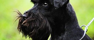
- Yorkshire terriers (almost 4/5 of all registered cases);
- chihuahua;
- Pomeranian and German Spitz;
- toy terriers;
- miniature poodles.
Did you know? The smallest Yorkshire terrier in the world weighed 113 g and had body dimensions comparable to a pack of cigarettes - 95 mm in length and 63 mm in height. This baby lived only 2 years, but was immortalized by being included in the Guinness Book of Records.
The second category of animals that also often exhibit respiratory stenosis are the so-called brachiocephalic breeds. We are talking about dogs that, in the process of selective selection and various crossings, have developed a non-standard skull shape - short, flat and flattened. Various breathing problems in such pets are almost a common condition. This group includes:
- pugs;
- Pekingese;
- bulldogs;
- Shar-Pei;
- boxers;
- Tibetan spaniels;
- griffons;
- Dogues de Bordeaux;
- bullmastiffs;
- Cane Corso;
- Shih Tsu, etc.
As for secondary collapse, it can develop in animals regardless of age and breed. This pathology is inherent in both small and large dogs; in addition, a similar problem also occurs in cats, cows and horses.
Why does ring flattening occur?
Primary weakening of the cartilage tissue of the rings is associated with low levels of glycosaminoglycans and glycoproteins. In ornamental pets it is considered a congenital anatomical defect associated with the likelihood of inbreeding.
Stent insertion (with accessory device)
Predisposing factors:
- respiratory, viral infections;
- incorrect tracheal intubation during anesthesia;
- neck injuries (including a sharp tug on the collar);
- allergies to odors, cigarette smoke, air fragrances.
More often, collapse is diagnosed in Yorkies, toy terriers, Pomeranians, and Chihuahuas. Symptoms appear in animals over 5 years of age spontaneously or when exposed to any factor. The breeder notices that the pet is coughing, its general condition is deteriorating, and “wheezing” (stridor) breathing appears. With severe stenosis, respiratory failure develops.
How do symptoms appear?
The clinical picture of tracheal collapse in the first stages of the disease is not always obvious. The first symptom that an owner needs to pay attention to is a cough without signs of a cold. The second is vomiting without signs of poisoning.
The peculiarity of cough as a sign of stenosis is its paroxysmal and dry (non-productive) form. As a rule, this occurs in response to quite characteristic stimuli, for example, a leash that is too tight, feeling the animal’s neck, or strong excitement that “tightens the throat.” Vomiting in this case is a consequence of overstraining the muscles of the larynx and has nothing to do with what the dog ate the day before.
Interestingly, cough can become not only a symptom of tracheal collapse, but also an indicator of it. For example, many acute respiratory diseases in dogs, primarily viral, are accompanied by a special kind of symptom, which has received a specific (unofficial) name - kennel cough. It happens that it is a strong cough caused by ARVI that manifests the previously asymptomatic narrowing of the tracheal rings, aggravates the pathology and thus makes it noticeable.
Did you know? When a dog gets tired, it loses its sense of smell. The fact is that for a speedy recovery, the animal needs to switch to breathing through the mouth, and in this case, the olfactory receptors perceive no more than 15% of the information.
In later stages of prolapse, its manifestations become more obvious. The owner may notice in his pet :
- shortness of breath (inspiratory, with difficulty inhaling, or expiratory, with difficulty exhaling);
- whistling and wheezing when breathing (sometimes with a characteristic pop from the mutual contact of the walls of the trachea);
- rapid fatigue, up to intolerance to any physical activity;
- cyanosis of the skin and mucous membranes;
- transition of cough to a productive form (expectoration of a large amount of sputum);
- suffocation.
Diagnostic process
If tracheal prolapse is suspected in a dog, the veterinarian can make an initial, presumptive diagnosis during a general examination of the animal. The problem is that palpation and palpation of the throat and neck in this case are very dangerous and provocative procedures, since they can cause a severe coughing attack, accompanied by spasms, vomiting and suffocation.
Therefore, to accurately determine the presence and stage of development of airway stenosis, special examinations are prescribed:
- X-ray of the cervical region (at the moment of inhalation) and the thoracic region (at the moment of exhalation) from the side;
- fluoroscopy (considered a more informative method compared to radiography, since it allows you to see the position of the trachea in dynamics, during the breathing process;
- endoscopy - tracheoscopy or tracheobronchoscopy (considered as the best diagnostic method in terms of visualizing the problem, since it allows you to reliably determine the stage of development of the pathology. It has one significant drawback - it requires general anesthesia);
- blood test (general and biochemical) - necessary when diagnosing secondary tracheal collapse to determine the causes of the problem;
- cytological and bacteriological sputum samples.
Important! If a dog has breathing problems, before tracheoscopy and after the procedure, the animal must undergo artificial ventilation - oxygen therapy.
Before starting the examination, the veterinarian must collect a detailed history: find out what the dog eats, what diseases it has suffered, etc. Depending on the answers received, additional types of tests and studies may be prescribed, for example, to identify concomitant pathologies, before total infectious, as well as endocrine and cardiovascular. This need is due to the fact that cough and difficulty breathing, in addition to tracheal collapse, are symptoms of many other diseases, so diagnosis by exclusion is very important for prescribing the correct treatment.
Diagnostic methods
In modern veterinary medicine, an effective method for diagnosing neoplasms is endoscopic examination. However, with the help of a special tube, the doctor can only determine the presence of a polyp. X-ray and ultrasound examination of the body will help make a differential diagnosis.
An accurate diagnosis is established based on histological examination of tissues. For this purpose, during endoscopy, a veterinarian performs a biopsy and removes a piece of the tumor for cytological examination.
How to relieve an attack
It is very important to detect the structure of your pet’s airways in time and thereby prevent them from completely closing. However, many owners are interested in a more specific question: what to do if the dog has already begun to choke, and there is simply no time to get to the veterinarian.
We have to admit that without special knowledge, drugs and equipment, it is almost impossible to help an animal with complete collapse of the trachea. In such cases, stenting may be necessary, i.e. installation of a special endotracheal device that provides the pet with the ability to breathe independently.
But usually such a condition does not develop unexpectedly, therefore, when talking about first aid during an attack, we usually mean a strong cough, accompanied by a reflex spasm of the larynx, when the animal is not even able to drink water. An effective method in this case is drugs containing codeine, an opium alkaloid used in medicine as a proven antitussive. The substance acts directly on those parts of the brain in which the “cough center” is located, as a result of which the sore throat disappears and spasmed muscles relax.
In practice, it is unlikely that it will be possible to relieve an attack in this way at home, since codeine, like any narcotic drug, cannot be purchased over the counter.
Did you know? The dog perceives the smell of drugs more strongly than the aroma of plant products. In countries where the transport of fruits and vegetables is prohibited, dogs can only help customs officers in finding carrots and apples, while these animals are simply irreplaceable in tracking drug traffickers.
Therefore, the only way to help your pet is to deliver it to the veterinary clinic as quickly as possible. On the way to the doctor, if the dog’s condition causes serious concern for its life, you can try to give the animal one of the milder and, accordingly, accessible sedatives designed to calm and relax the patient.
How is tracheal collapse treated in dogs?
Tracheal collapse can be treated in two ways: conservative (with the help of medications) and operative (surgical). The decision in each specific case is made by the doctor, depending on the degree of narrowing of the lumen in the respiratory tract, the general condition of the animal, its age and other individual factors.
With the help of drugs
If airway stenosis is at the first or second stage of development, the animal can be helped without surgery. Such treatment, however, does not provide recovery, but simply helps to relieve the clinical manifestations of the pathology, i.e. it is symptomatic.
Important! Conservative treatment is justified for secondary tracheal collapse, since in this case there is hope that after eliminating the causes that led to the development of the pathology, the problem will be completely resolved.
In addition to the use of medications, during the stabilization stage it is very important to provide the dog with complete rest and limit its mobility as much as possible in order to prevent the occurrence of factors that could provoke an attack.
Taking into account all available indications and contraindications, the doctor can prescribe the following groups of medications to the animal:
| Category of veterinary drug | Examples | Indications for use |
| Antibiotics | Amoxicillin, Ceftriaxone, Ciprovet, Marfloxin, Baytril | Used to identify a bacterial infection causing coughing attacks |
| Antiviral agents | Phospenyl, Neotime, Anandin, Dostim, Camedon, Neoferon | Accelerate the process of stopping a respiratory viral infection if it aggravates the dog’s condition |
| Glucocorticoids | Dexamethasone, Hydrocortisone, Triamcinolone, Betamethasone | Block inflammatory processes that provoke coughing |
| Bronchodilators | Ventipulmin, TheoDur, Slo-Bid gyrocaps | Expand the airways and relax the tracheal muscles |
| Mucolytics | Mucoltin, Bronchipret, Butamira, Terpinhydrate | Facilitate the discharge of sputum, thereby making attacks less painful and shorter |
| Antitussives | Hydrocodone | Affects the brain, disabling the “cough center” |
| Antispasmodics | Deepprovide, ZooHealth | Relaxes muscles, relieves coughing |
If the use of the listed remedies does not help or has too little effect, the veterinarian decides to perform an operation.
Carrying out the operation
The good news is that, according to statistics, surgical treatment of tracheal collapse is necessary in no more than 7% of cases. Usually we are talking about the third and fourth stages of the disease, when the lack of a radical solution to the problem can lead to the development of complete stenosis, which, in turn, can be fatal. It is important to understand that drug therapy is often not canceled, but supplemented by surgery, although it is surgical intervention that provides the maximum effect of treatment.
There are several methods of surgical treatment of tracheal collapse, but the most common are two types of stabilization - intraluminal and extraluminal, as well as creation of a fold of the dorsal tracheal membrane.
In the latter case, after making an incision in the trachea, the surgeon suturing the membrane, thus eliminating its sagging. The method is justified in cases where the pathology is insignificant. In later stages of the disease, suturing can be dangerous because it causes even more severe narrowing of the lumen.
The essence of surgical intervention in the case of intraluminal stabilization is the installation of a self-expanding stent in the most narrowed lumen of the trachea due to pathology - a small nitinol (nickel and titanium alloy) tube made of a fine mesh. Such a stent does not allow the airways to completely close, expanding the lumen by about one and a half times and thereby guaranteeing the animal’s life. This treatment method appeared relatively recently, but is already showing very good results. Its main advantage is its relative simplicity and minimal trauma: the entire procedure takes no more than 10 minutes. Similar stents are also used in general surgery (though they are placed in people for other indications), so it is reliably known that some time after the operation the mesh completely adapts to the site of its installation, and the patient does not feel any discomfort.
External luminal stabilization is the classic surgical treatment for tracheal collapse in dogs. It involves installing polypropylene rings into the trachea, and even a tube cut across from a disposable plastic syringe can serve as an “implant.” To maintain the flexibility of the fabrics, the plastic stabilizer is not a full circle, but approximately 2/3 of its part. The operation is considered quite risky, since an inexperienced surgeon may touch nerves or blood vessels during the procedure, which is fraught with serious complications, primarily necrosis of the tracheal walls.
Did you know? Tracheostomy is by far the most expensive operation in the world. Its price in some clinics can reach $
.
How to care for your dog after surgery
Postoperative care for the dog directly depends on what method of surgical treatment was used. This period lasts from several days to one and a half months and includes the mandatory implementation of the following activities:
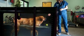
- oxygen therapy - using oxygen concentrators or, in severe cases, artificial ventilation;
- a course of antibiotics for 7-10 days - to prevent a bacterial infection that could be introduced into an open wound during surgery or during the healing stage;
- the use of corticosteroids, anti-inflammatory, antitussive drugs, as well as bronchodilators - in order to relieve possible coughing attacks, which often occur during the period of implant engraftment, including due to swelling of the upper respiratory tract;
- if indicated, follow-up examinations with a cardiologist (such precautions are necessary if there is reason to suspect collapse of the main bronchi or heart failure);
- a therapeutic diet that includes dietary feed, as well as vitamin and mineral supplements that stimulate the immune system.
If rehabilitation is difficult and is associated with difficulty breathing, sometimes it is necessary to resort to extreme measures - tracheostomy, i.e. repeated surgical intervention aimed at creating an opening in the trachea to provide air access to the lungs.
Diagnosis of the problem
The level of modern diagnostic medicine allows us to easily identify this disease.
A number of studies and tests are used to identify polyps:
- Ultrasound of the genitourinary tract,
- cystoscopy, cystography,
- general and detailed urine analysis,
- biopsy,
- MRI.
Also, to make a diagnosis in each specific case, other diagnostic measures can be taken to clarify or exclude certain pathologies.
Prognosis and possible complications
If the operation is performed on time and performed by an experienced surgeon, the prognosis is regarded as very favorable. The result of treatment is usually assessed by control bronchoscopy, which is performed on average one and a half months after stenting or another method of surgical stabilization.
However, a lot depends on the owner himself. Until the animal’s health status is completely back to normal, it is extremely important to protect the pet from any adverse effects of the external environment: infections, stress, injuries, etc. Any chronic diseases that the dog suffers from should also be identified and monitored. In addition, no matter how high-quality the implant is, we are still talking about a foreign body, so the process of its engraftment should also be carefully monitored. For this, if necessary, endoscopic and fluoroscopic examinations are carried out, which, in case of the slightest doubt about the pet’s well-being, its owner must insist on.
Important! Installing a stent or implant is not a treatment for the disease. We are talking only about mechanically ensuring the normal breathing process by maintaining the necessary lumen in the trachea.
Possible postoperative complications include:
- tissue necrosis;
- allergic reaction to the material from which the stent is made;
- displacement (migration) of the stent;
- implant deformation;
- persistent cough that cannot be relieved with any medications;
- pathological growth (hyperplasia) of the tracheal mucosa.
Even after a successful operation, the dog will still cough, since the implant itself is an irritant to the mucous membranes.
What happens to the dog after surgery
If the owner chose to undergo surgery , then after it it will be necessary to carry out therapy for the best recovery. Constant monitoring by a veterinarian, selection of medications to prevent infection, as well as chronic complications, will be mandatory. The owner must strictly follow the doctor’s instructions and recommendations for evacuating mucus from the dog’s body, since the stent and implant do not always allow the animal to cough up mucus on its own.
From time to time it is worth taking your dog to the veterinarian for an endoscopy and x-ray examination to closely monitor the implant or stent.