What is dangerous about enlarged submandibular lymph nodes in a dog?
Even a slight increase in the submandibular lymph nodes in a dog should not be ignored. This is especially true for animals over the age of 5 years. It is during this period that the largest number of tumor processes are diagnosed. At the same time, the pet may seem absolutely healthy in appearance, without giving up food and its usual lifestyle. In young individuals, this symptom may indicate problems in the nasopharynx. Well, most often the reason lies in dental problems, which can occur even in puppies.
Is an abscess dangerous?
An abscess is a formation filled with pus; many also call it an abscess. The growth occurs at the site of a bite, chemical, thermal or mechanical damage to the skin. After the abscess is fully matured, it is removed. Only a veterinarian should do this if you do not want to harm your animal. If the abscess is opened untimely or incorrectly, the dog may develop severe complications and die.
To make the growth mature faster and stop bothering the animal, you can apply heat to it. Never put pressure on the abscess or pick at it. When removing a formation, great care is taken so that the infection does not enter the blood. After the operation, the wound is disinfected with iodine solution. Keep the wound open to help it heal faster, treat it with antiseptics every day.
Description of possible diseases
If the dog's lymph node under the jaw is enlarged, you need to carefully examine the oral cavity for the appearance of abscesses, fistulas on the gums, and caries or tartar on the teeth. Be sure to also palpate the remaining lymph nodes (under the arms, neck, groin). If there are signs of inflammation, then it is better not to hesitate to visit the veterinarian. It is worth keeping in mind the following diseases:
- Mycoplasmosis. Often found in puppies under one year of age. It almost always occurs against the background of hidden diseases (rhinitis, follicular conjunctivitis). There may be discharge from the eyes, watery eyes, swelling of the muzzle, and enlarged submandibular lymph nodes. Any veterinarian will tell you that when taking a smear, even a healthy dog can find mycoplasma cells. But, if there are symptoms, then they usually resort to taking sumamed, doxycycline and clindamycin, to which mycoplasma is sensitive. In most cases, symptoms subside within 3-5 days. If this does not happen, then the cause must be sought in other, more serious diseases.
- Juvenile cellulite. It most often affects puppies between two weeks and several months of age. It always occurs with different clinical signs. Some people have swollen muzzles and sour eyes, while others suffer from deep skin lesions in the nose and lips. From a general point, it can be noted that in almost all puppies the submandibular lymph nodes become enlarged. When palpated, they feel like peas and are not accompanied by pain. In some cases, the ears and cervical lymph nodes may be involved in the process (they also increase in size). Therapy is selected individually, depending on the severity of the clinical picture. To exclude bacterial damage, cytology of the fluid from the inflamed areas is performed. There are many known cases where juvenile cellulitis went away on its own, but some owners and veterinarians do not want to risk the health of their pet and do not expect spontaneous recovery. As treatment, injections of prednisolone are used at the rate of 1 mg per 1 kg per day.
- Deep periodontal disease. Severe gum damage that can lead to partial or complete loss of teeth. Occurs in dogs after 7 years of age. Some breeds have a predisposition to this disease due to the structural features of the muzzle and malocclusion (Yorkies, bulldogs, pugs). The reason may lie in tartar or deep caries. At the initial stage, the gums turn red and become loose. As the process progresses, the submandibular lymph nodes enlarge and bleeding from the gums appears. The pet experiences pain while chewing food, which is why over time it may begin to refuse food. Periodontal disease is difficult to treat, especially in advanced forms, since in some cases it is impossible to do without removing all the teeth. The age of the animals complicates the situation, since the removal procedure is carried out under anesthesia. In some cases, significant improvements can be achieved through oral sanitation, treatment of carious teeth and stone removal.
- Abscess. It can form on the gum or tooth root. In the first case, you can immediately notice the appearance of a growth on the gum and help your pet in time, take him to the dentist. In the case of the formation of an abscess on the root of a tooth, everything is much more complicated, since something is wrong can be noticed only after the submandibular lymph nodes are enlarged, the cheek or the entire muzzle is swollen. An abscess can be caused by a foreign body entering the gum, caries and dental plaque. Abscesses on the gums are opened, cleaned of pus and treated with an antiseptic. In case of damage to the tooth root, the option of removal is chosen.
- Lymphosarcoma. The most dangerous cause of enlarged submandibular lymph nodes in a dog. Occurs mainly in old individuals. At the initial stage, you can feel two peas under the jaw. There is usually no pain. As the process progresses, the cervical, axillary and inguinal lymph nodes enlarge. The animal begins to swell. If the process spreads to the bone marrow and internal organs, then this is considered the last stage of lymphosarcoma. Chemotherapy may be used as treatment. Surgical intervention is considered ineffective, since there is a risk of the process spreading to other lymph nodes. With the help of drug therapy, remission can be achieved (up to 5 months). But, you need to understand that with lymphosarcoma the prognosis is almost always unfavorable.
Symptoms
The listed types of growths on the gums in dogs are united by a set of similar symptoms:
- an unpleasant odor is felt from the mouth, sometimes slightly putrid;
- There may be traces of blood or pus on the food bowls;
- the gums at the site of the lesion are swollen, edematous, reddened;
- salivation (salivation) is increased, especially during meals;
- appetite is reduced in the form of pain when chewing;
- the temperature is elevated;
- teeth are loose and can sometimes fall out;
- withdrawn, lethargic behavior;
- when touching a painful area, the pet shows aggression;
- With a hard cancerous growth on the gum, the dog may experience emaciation.
These symptoms do not always indicate the appearance of neoplasms. They can be indicators of caries, tartar, and the appearance of wounds in the oral cavity.
Enlarged submandibular lymph nodes in a dog
Finally, I would like to remind you that the dog’s submandibular lymph nodes are normally palpable, but they should not hurt or increase in size (a pea is already considered a sign of inflammation).
source
Jaw tumor in dogs
Oral tumors are quite common in dogs. As a percentage, they occupy more than 99% of all tumors of the gastrointestinal tract and 11-15% in the structure of all tumors. Males and young animals are more predisposed to their occurrence.
Content
Localization of tumors
Most often, the tumor is localized in the jaw area of a dog (more than 75% of all oral tumors), followed by the lips (17%), and less commonly, the tongue (8%).
Causes
Among the possible causes, more attention is paid to canine and feline papilloma viruses, since it is believed that the development of papillomas and squamous cell carcinoma of the oral cavity is associated with them.
Symptoms
Symptoms of the disease include excessive salivation, sometimes with blood, bad breath, difficulty swallowing food, bleeding from the mouth, weight loss, tooth loss, growths in the mouth, enlarged lymph nodes, swelling around the eyes.
Diagnostics
To diagnose jaw tumors, radiography, biopsy, magnetic resonance imaging (MRI), and computed tomography (CT) are performed. The choice of diagnostic method is determined by the doctor. Instrumental diagnostics of the affected areas helps to assess the size of the tumor and, depending on this, determine methods of its treatment. Approximately 60-80% of gum growths penetrate into the bone tissue of the jaws, this is clearly visible on x-rays, MRI and CT. The tumor can affect both bone tissue (22%) and soft tissue (78%).
Types of benign tumors
The most common benign tumors are:
Odontoma. This tumor formation of odontogenic origin is the result of improper formation and development of the tooth embryo. If it is large, it becomes infected. In these cases, there are signs of osteomyelitis and fistula formation. Most often localized on the lower jaw, especially in the molar area. Treatment is surgical.
Adamantinoma. In most cases, adamantinoma (ameloblastoma) is localized in the lower jaw and is much less common in the upper jaw. Adamantinoma (ameloblastoma) is a benign epithelial tumor, its structure is similar to the structure of the tissue of the enamel organ of the tooth germ. .The treatment is surgical.
Epulis. Clinically, it occurs as a slowly growing tumor formation on the buccal or (less often) lingual side of the gum. Radiologically, epulis is a benign periodontal growth.
Osteoma. A compact osteoma occurs on the upper jaw. Osteoma often occurs in the air cavities of the skull and on an x-ray image appears as a dense, structureless shadow.
Fibroma. It is also relatively common. It produces a round defect without any special radiological signs.
Types of malignant tumors
The most common malignant tumors are:
Malignant melanoma. The tumor is more common in animals with pigmented black skin and coat (black terriers, giant schnauzers, etc.). This neoplasm has a tendency to metastasize to regional lymph nodes, lungs, and brain. Treatment is surgical excision of the tumor in combination with radiation therapy, sometimes cryodestruction.
Fibrosarcoma. This is an infiltrating tumor that metastasizes in less than 20% of cases. Often recurs after surgical excision. The main method of treatment is excision of the tumor within healthy tissue.
Squamous cell carcinoma. The tumor rarely metastasizes. Regional chemotherapy or chemoradiotherapy may be an alternative to surgical excision. In dogs, the treatment of choice is surgery. However, if the tumor is difficult to remove, the animal is given radiation therapy or chemoradiotherapy in the preoperative period.
Ameloblastoma. The tumor destroys soft tissue and bone at its location. Does not metastasize. Treatment is surgical removal within the healthy bone. To determine the boundaries of the tumor, an x-ray, CT or MRI are performed, all of which help determine the boundaries of the tumor.
Osteosarcoma. Tends to grow into various tissues of the jaw apparatus. Treatment is surgical. Proper removal of osteosarcoma can not only save the life of the animal, but also provide a good level of quality of life.
The article was prepared by Milyushkina E.N., veterinary surgeon, endoscopist "MEDVET" © 2021 SEC "MEDVET"
source
Why does a tumor appear on the neck of dogs?
Benign formations under the skin:
- Hematomas. A soft formation occurs due to disruption of the integrity of blood vessels due to injury. In rare cases, subcutaneous hemorrhages may be painful to palpation.
- Lymphoextravasate. Severe trauma can lead to rupture of not only blood vessels, but also lymphatic ones, which is accompanied by the accumulation of lymph under the skin in the form of a lump.
- Abscess. A bite from a relative, a puncture wound, or a deep splinter are often complicated by a bacterial infection, resulting in swelling. Violation of the rules of asepsis and antiseptics during injections leads to the development of an inflammatory process.
- Insect bites (bees, wasps, horseflies).
- Warts, papillomas . Such bumps do not cause concern to the pet and are typical for short-haired individuals. As a rule, the growths have a brown color and a heterogeneous (lumpy) surface. Papillomas resemble cauliflower, soft and loose in consistency. The owner should be alert to a wart or papilloma from which blood is oozing.
- Purulent skin diseases (pyoderma). The bumps are caused by the development of a bacterial infection. Pyoderma bumps are found not only on the neck, but also quickly spread throughout the pet’s body.
- Cyst . A cystic formation on the neck of dogs is a rare occurrence and is formed when the ducts are blocked or compressed.
In some cases, the owner may mistake an enlarged peri-cervical or submandibular lymph node for a neoplasm.
- Lipoma . A benign neoplasm under the skin is a ball-shaped compaction and consists of adipose and connective tissue. The lump usually occurs in dogs over 5 years of age.
- Hemangioma, keratoacanthoma, fibroma . Neoplastic tumors such as trichoepithelioma and pilomatrixoma are less commonly diagnosed. Typical for animals older than 5-6 years. In some cases, there is a breed predisposition.
Malignant tumors in dogs include primarily hemangiosarcoma, melanoma, liposarcoma, and fibrosarcoma. Mast cell tumors develop quickly and are characterized by infiltration into other tissues and the development of metastases. The general condition of the four-legged friend worsens. The dog loses its appetite, becomes lethargic and inactive.
What to do if detected:
- it is necessary to examine the pet for pain and signs of inflammation;
- You should not self-medicate, but visit a specialized institution and find out the nature of the neoplasm.
Diagnosis of an animal includes:
- thorough examination of the animal, palpation
- biopsy - taking biological material followed by bacteriological or cytological examination;
- general and biochemical blood test;
- X-ray examination, computer and magnetic resonance imaging.
Treatment of a tumor on the neck in dogs:
- Inflammatory formations on the pet's neck can be successfully treated with the use of antibacterial drugs.
- In some cases, a veterinarian resorts to opening the abscess.
- Papillomas and warts are removed.
- Cancerous tumors are removed surgically; laser methods of treating the tumor and chemotherapy are also used.
Read more in our article about tumors on the neck in dogs, causes and treatment.
Types of neoplasms
All lumps found in dogs can be divided into groups: non-cancerous formations and malignant (cancerous) tumors.
Non-cancerous lumps:
- Papillomas and warts are found mainly in smooth-haired dogs. According to scientists, the cause of the formations may be a viral infection existing in the pet’s body. Warts appear in the form of a brownish growth of the dermis. Most often, they do not cause pain; consultation with a veterinarian can only be recommended for preventive purposes.
- Cysts. They can appear anywhere on an animal’s body, including the chin. Most often, swelling is discovered by chance, and not during a routine examination. When the seals are localized on the pet's head or face, they are difficult to miss.
- Hematomas. The appearance of lumps caused by hematomas is most often observed in the postoperative period, especially when blood vessels are damaged. These bumps are soft to palpation and can change the shape of the area of the body where they are located. Most often, hematomas do not cause pain, but the animal may experience discomfort associated with them.
- Insect bites. Bumps caused by insect bites are noticeable on your pet's face and chin.
- Abscesses. The formation of abscesses is caused by the penetration of bacteria into the body. The development of an abscess is most likely after bites and puncture wounds. An abscess is characterized by inflammation of the subcutaneous layer of the epidermis and a rise in temperature; these manifestations are accompanied by pain sensitivity. The formation time can take up to several days. There may be discharge of purulent contents.
Tumor formations are divided into two groups:
- Benign neoplasms - their distinctive feature is the absence of metastases and limited localization in the body. Typical lumps can reach large sizes; therefore, most owners insist on removing benign tumors;
- Malignant lumps can metastasize (spread), destroying surrounding tissues and organs. The formations can provoke bleeding due to rupture of the dermis.
Skin seals may not cause significant discomfort to the animal and remain unchanged in size for a long time. The size and density of formations may vary and depend on the origin and histological structure.
Types of bumps on the neck
When caring for a pet, while playing, the owner may notice a tumor on the dog’s neck. There is no need to panic, since in most cases the neoplasms are not associated with a malignant course and can be treated with medication or surgery. According to veterinary experts, lumps in the neck area of four-legged pets can be of two types – those not associated with cancer and those with malignant tumors.
Benign formations under the skin
Benign neoplasms localized near the pet’s neck include the following types of lumps:
- Hematomas . Mobile, active and inquisitive animals often get injured. Soft formation occurs due to a violation of the integrity of blood vessels. In rare cases, subcutaneous hemorrhages may be painful to palpation.
- Lymphoextravasate . Severe trauma can lead to rupture of not only blood vessels, but also lymphatic ones, which is accompanied by the accumulation of lymph under the skin in the form of a lump.
- Formations on the neck can also be of an inflammatory nature, such as an abscess . A bite from a relative, a puncture wound, or a deep splinter are often complicated by a bacterial infection, resulting in swelling. Violation of the rules of asepsis and antisepsis during injections leads to the development of an inflammatory process, accompanied by the formation of a dense swelling.
- An abscess is usually accompanied by an increase in local and general temperature and pain. Often the dog loses its appetite, becomes lethargic and inactive.
Abscess
- Insect bites . In the summer, four-legged pets can be attacked by bees, wasps, and horse flies. At the site of the bite, the owner discovers a lump.
- Warts, papillomas . Viral growths are a common type of benign tumor on the neck in dogs. Such bumps do not cause concern to the pet and are typical for short-haired individuals. As a rule, the growths have a brown color and a heterogeneous (lumpy) surface. Papillomas resemble cauliflower, soft and loose in consistency. A wart or papilloma from which blood is oozing should alert you.
- Purulent skin diseases . Pyoderma is often characterized by the formation of specific bumps under the skin caused by the development of a bacterial infection. Pyoderma bumps are found not only on the neck, but also quickly spread throughout the pet’s body.
- Cyst . Cystic formations on the neck of dogs are not a common occurrence. However, when the ducts are blocked or compressed, a soft and loose, usually painless lump may develop on the neck.
In some cases, the owner may mistake an enlarged peri-cervical or submandibular lymph node for a neoplasm.
And here is more information about the causes of goiter formation in dogs.
Tumors on the neck, under the jaw
In veterinary practice, quite often, when an owner contacts him about a lump on the neck, a lipoma is discovered in a four-legged patient. A benign neoplasm under the skin is a ball-shaped compaction and consists of fatty and connective tissue. The lump usually occurs in dogs over 5 years of age. Breeds such as Labradors and Retrievers are predisposed to the pathology. Lipomas are often observed in cocker spaniels.
The owner most often finds a single formation on the pet’s neck. In the practice of veterinarians, there are cases when the lipoma has a symmetrical localization. A benign neoplasm has virtually no effect on the general condition of the pet. The dog has retained its appetite, there is no lethargy or depression.
The owner's concern is appropriate if the wen causes inconvenience to the pet, has grown to a decent size and interferes with walking and running.
In addition to lipoma, benign neoplasms on the neck in dogs also include gamangioma, keratoacanthoma, and fibroma. Neoplastic tumors such as trichoepithelioma and pilomatrixoma are less commonly diagnosed.
Neoplasms, according to veterinary experts, are typical for animals older than 5-6 years. In some cases, there is a breed predisposition. Thus, in German shepherds, keratoacanthoma is often observed, which is limited small nodules.
Pilomatrixoma, often found in terriers and poodles, is a cystic tumor. The size of the neoplasm can reach 10 cm in diameter. Tricholemmoma on the neck, common in Afghan hounds, can grow up to 17 cm in diameter.
Malignant tumors in dogs include primarily hemangiosarcoma, melanoma, liposarcoma, and fibrosarcoma. Mast cell tumors develop quickly and are characterized by infiltration into other tissues and the development of metastases. The general condition of the four-legged friend worsens. The dog loses its appetite, becomes lethargic and inactive.
Possible diseases
Now let's look at the options in which you should never delay visiting the veterinarian if you feel even a soft lump on your dog's chin:
Characteristic features: a hard cone, prone to rapid growth. If it forms under the jaw, problems with chewing and swallowing may occur, hence the animal’s refusal to eat.
At the initial stage, it is successfully treated surgically, provided there are no metastases. Pets over 10 years old or with advanced cases may be denied surgery, as there is a high risk that the dog will not be recovered from anesthesia.
It is characterized by a rapid course, large swelling and acute pain at the site of the lesion. It may be necessary to open the tumor to drain the pus. A drainage is also applied to drain any remaining pus and blood.
Regular treatment of the oral cavity with antibacterial drugs is indicated. After removing the inflammation, they take on the very reason for the appearance of gumboil - they treat the gums or teeth.
Common among dogs of all ages. Most often they appear on the face (eyelids, chin, cheeks).
Each formation is subject to careful examination by a veterinarian, since papillomas have a tendency to transform into cancerous tumors! At the initial stage, single growths can be felt like a small pimple. In some cases they grow to the size of a large pea.
When injured, they bleed and take a long time to heal. The need for removal is determined by a veterinarian. For example, in English and French bulldogs, papillomas are an invariable attribute of the breed. They have several dozen of them.
A benign formation that is soft to the touch and is not accompanied by pain. It can grow for several years before its owner notices it.
In some cases, tumors reach the size of a chicken egg. All lipomas are removed surgically. There is no tendency to develop a malignant process. More common in older dogs.
You also cannot ignore the enlargement and inflammation of the lymph nodes, which can also provoke the appearance of solid tumors on the dog’s chin. This process may indicate serious problems with the thyroid gland.
To post a reply you must log in or register.
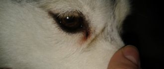
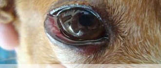
Hello. About a month ago, the dog's eye became inflamed (there was purulent discharge), and after some time a red-burgundy lump appeared in the inner corner of the eye. The hospital said it was an adenoma of the third eyelid and suggested surgery. Literally a few days after the appearance of this adenoma, the dog’s right jaw began to rattle literally overnight. I don't know if this is related. Then they prescribed Biseptol and Gammavit. Three days later the swelling was almost gone. A week later they had surgery. Just yesterday history repeated itself again. The dog has swelling under the right jaw from below, on the very lower jaw there is like a tumor, hard and hot to the touch. Viscous saliva flows from its mouth, but the dog does not refuse food and is relatively cheerful. Our veterinarians did not find a connection between the eye and this tumor. They say she may have caught a cold. In fact, these two days it was cold and raining. The dog lives outside and could easily have become hypothermic. Maybe this is important. After surgery to remove the adenoma, blood flowed from the eye for quite a long time (at least 2 hours). Those. We stopped her, as soon as the dog moved, the stream started flowing again. Maybe the clotting is bad? The dog sits on a chain with a collar. Jumps a lot, maybe he hurt himself? Tell me what this could be? There is little hope for our veterinarians. The dog is not mine, my younger brother’s. I can go twice a day. Can you suggest a treatment? Does it make sense to install gammavit again? They prescribed tablets - broad-spectrum antibiotics. I haven’t seen it myself yet, but according to my brother it’s called sulfite. 480 mg. tablet. The dog is a male. Soon it will be 2 years. Weight 36 kg. Metis of a Saint Bernard woman and her half Saint Bernard son. These are my dogs, and it is my fault that such puppies appeared.
Hello. The connection between discharge from the eye and swelling of the jaw can be direct - a bad tooth! A thorough examination is needed; the dog may have pulpitis; severe inflammation often sets in and, as a result, conjunctivitis occurs. Also, inflammation can spread to the jaw itself and spread to other teeth. Naturally, if all the changes are on one side (you did not specify which eye was affected). The affected tooth must be removed. Such operations are performed under general anesthesia. Then a course of antibiotics and local treatments is carried out. Another possibility is inflammation of the trigeminal or facial nerve.
Thank you very much for your answer. Unfortunately, the treatment that was prescribed to us did not help at all (the doctor also suspected an allergy), yesterday morning I took it home, when I came home from work, I saw that the tumor was beginning to cover the other half of the muzzle, I realized that I had to pull and treat the dog for Allergies no longer make sense. I made an appointment at a private clinic in the regional center. There they did an X-ray and came to the conclusion that it was an abscess. They opened it up under general anesthesia, found it and cleaned it. Now he has drainage tubes and gauze in his cheek. Adenoma, discharge from the eye and tumor were on one side. The doctor said that the inflammation was due to the lymph node. Does this mean he is hypothermic? If possible, answer, would it be visible on the X-ray photographs if the tooth is inflamed or broken? The surgeon examined his mouth briefly, this was when he was already under anesthesia, and said that everything was fine with his teeth and gums. When the surgeon performed the operation, he injected some kind of drug, white, like milk, with a strong chemical smell. The operation lasted a long time, an hour and a half, two. He literally injected 3 cubes into a vein every 15 minutes, where an IV with saline solution was placed. He only said that it was not a drug, but the dog would sleep and not feel anything. Is this drug very harmful to the liver/kidneys? If, of course, he can be identified from my story.
Hello. How is your dog feeling now? An abscess is a local inflammation of tissue that occurs due to external trauma, if an infection occurs, or due to an internal cause, such as a tooth or lymph node. The damage to the tooth would be visible on an x-ray, as well as during an examination; enlargement and inflammation of the lymph node are also visible upon examination. Where exactly was the abscess located, in the skin, cheek muscles, inside the gum? I find it difficult to say what drug the surgeon administered.
A tumor is swelling - a natural reaction. How is the dog feeling now?
Benign formations under the skin:
- Hematomas. A soft formation occurs due to disruption of the integrity of blood vessels due to injury. In rare cases, subcutaneous hemorrhages may be painful to palpation.
- Lymphoextravasate. Severe trauma can lead to rupture of not only blood vessels, but also lymphatic ones, which is accompanied by the accumulation of lymph under the skin in the form of a lump.
- Abscess. A bite from a relative, a puncture wound, or a deep splinter are often complicated by a bacterial infection, resulting in swelling. Violation of the rules of asepsis and antiseptics during injections leads to the development of an inflammatory process.
- Insect bites (bees, wasps, horseflies).
- Warts, papillomas. Such bumps do not cause concern to the pet and are typical for short-haired individuals. As a rule, the growths have a brown color and a heterogeneous (lumpy) surface. Papillomas resemble cauliflower, soft and loose in consistency. The owner should be alert to a wart or papilloma from which blood is oozing.
- Purulent skin diseases (pyoderma). The bumps are caused by the development of a bacterial infection. Pyoderma bumps are found not only on the neck, but also quickly spread throughout the pet’s body.
- Cyst. A cystic formation on the neck of dogs is a rare occurrence and is formed when the ducts are blocked or compressed.
In some cases, the owner may mistake an enlarged peri-cervical or submandibular lymph node for a neoplasm.
- Lipoma. A benign neoplasm under the skin is a ball-shaped compaction and consists of adipose and connective tissue. The lump usually occurs in dogs over 5 years of age.
- Hemangioma, keratoacanthoma, fibroma. Neoplastic tumors such as trichoepithelioma and pilomatrixoma are less commonly diagnosed. Typical for animals older than 5-6 years. In some cases, there is a breed predisposition.
Malignant tumors in dogs include primarily hemangiosarcoma, melanoma, liposarcoma, and fibrosarcoma. Mast cell tumors develop quickly and are characterized by infiltration into other tissues and the development of metastases. The general condition of the four-legged friend worsens. The dog loses its appetite, becomes lethargic and inactive.
What to do if detected:
- it is necessary to examine the pet for pain and signs of inflammation;
- You should not self-medicate, but visit a specialized institution and find out the nature of the neoplasm.
Diagnosis of an animal includes:
- thorough examination of the animal, palpation
- biopsy - taking biological material followed by bacteriological or cytological examination;
- general and biochemical blood test;
- X-ray examination, computer and magnetic resonance imaging.
Treatment of a tumor on the neck in dogs:
- Inflammatory formations on the pet's neck can be successfully treated with the use of antibacterial drugs.
- In some cases, a veterinarian resorts to opening the abscess.
- Papillomas and warts are removed.
- Cancerous tumors are removed surgically; laser methods of treating the tumor and chemotherapy are also used.
Read more in our article about tumors on the neck in dogs, causes and treatment.
Diagnosis of an animal
When an owner contacts you about a dog having a tumor on its neck under the skin, a veterinarian will first of all conduct a thorough examination of the animal. The appearance, nature of the neoplasm, its size and consistency, and structure will help to identify the nature of the lump. By palpation, the doctor will determine the presence of pain in the pet, as well as the consistency of the tumor.
A diagnostic method such as a biopsy may be informative. Taking biological material followed by bacteriological or cytological examination will shed light on the nature of the tumor in a furry patient. In some cases, a veterinarian may recommend a general and biochemical blood test to clarify the diagnosis.
The veterinary therapist also has an X-ray examination, which can be used to determine the neoplastic nature of the tumor and identify metastases in the malignant course of the process. Equipped modern clinics also use computer and magnetic resonance imaging for diagnostic purposes.
Treatment of a tumor on the neck in dogs
If a tumor is found in a dog’s neck under the jaw, treatment directly depends on its cause. Inflammatory formations on the pet's neck can be successfully treated with the use of antibacterial drugs. In some cases, a veterinarian resorts to opening the abscess.
Attention should also be paid to the treatment of warts and papillomas. As a rule, professionals recommend removing these tumors. Firstly, the growths can be injured when the pet walks and rests, and secondly, one should not discount the possibility of such lumps degenerating into malignant structures.
With the help of a surgical scalpel it is possible to stop a cancerous tumor in a dog. Modern veterinary medicine significantly prolongs life and improves its quality when a terrible diagnosis is established. To treat a dog, laser methods are used to target the tumor, as well as chemotherapy.
And here is more information about the causes and treatment of lymphadenitis in dogs.
A tumor on the neck of a four-legged family member requires the owner not to delay a visit to a specialized institution. A veterinary specialist will determine the nature of the neoplasm and determine a therapeutic strategy. In some cases, a furry patient will need surgery.
What diseases can manifest themselves in this way?
A growth on the gum of a dog can be the main diagnosis of a disease, or it can only accompany it as a coincidence. If you find something like this in your pet’s mouth, you need to be prepared that the veterinarian can diagnose one of the following diseases:
- Papillomatosis. Simply put, these are warts. They do not pose any particular danger, but they occur against the background of a weakened immune system. That is, a caring owner should reconsider the lifestyle of his pet. Papillomas in dogs are not dangerous, but they can significantly interfere with leading an active lifestyle. They are usually removed with the help of medications, but if this is impossible or it turns out that their size and location make it difficult to eat normally, then they can also be removed surgically.
- Abscess. To immediately put an end to possible misconceptions, it should be said that an abscess is not a pimple or a boil. This is a purulent formation that occurs at the site of open wounds. Under no circumstances should you open it yourself, even if you are very confident in your own abilities. Improper opening of an abscess can lead to serious complications, possibly even blood poisoning. The veterinarian will also determine the origin of the abscess in the dog's mouth, which will allow him to make recommendations on how to avoid its reoccurrence.
- Hematoma. It is the result of a one-time severe injury or a consequence of constant mechanical impact on the same place. Small hematomas resolve on their own when any impact on the affected area stops. Large ones may require surgical removal.
- Cyst. It is essentially a benign tumor filled with fluid. It grows slowly and does not always require immediate surgical removal.
- Benign tumor. They can reach very significant sizes because they do not disturb the animal in any way. Veterinarians often recommend surgical removal, since the prognosis in this case is favorable - the tumor should not reappear.
- Cancer. Malignant tumors are also removed surgically, but this is only part of the treatment. Many research methods and chemotherapy are used. The danger of cancer in a dog’s mouth is the extremely rapid spread of metastases to the stomach and lungs. Therefore, in diagnosis and observation, not only laboratory tests are used, but also x-rays and ultrasound.
- Ulcerative stomatitis. In this case, the growth is an ulcer, often soft and suppurating. The appearance of ulcerative stomatitis indicates a significant violation of food hygiene and teeth cleaning in a dog.
The bulges and growths seen on the jaws do not always indicate any serious diagnosis. They may be the result of injury or temporary dental problems. You should be concerned if a hard growth appears on your dog's gum - it may turn out to be a tumor. Self-diagnosis is useless here; you must immediately contact a veterinarian.
Swelling of the face in dogs: etiology, symptoms, treatment
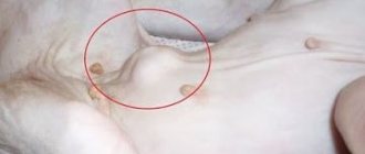
Many dog owners are faced with this problem when their pet’s limbs, lips, ears, muzzle suddenly swell, mainly in the eye area, lower jaw, and neck. The development of edema occurs due to excessive accumulation of fluid (transudate) in the subcutaneous layers, which is explained by an increase in the porosity of the vascular walls.
Swelling of the dog's muzzle is the most common pathology that occurs due to the development of angioedema (Quincke's edema) . If, in addition to swelling, the epidermis is covered with small red blisters, this indicates the development of urticaria (urticaria). Despite the fact that the animal’s condition may return to normal after a few hours or days, it is better to immediately seek help from a veterinarian.
Etiology, causes of swelling
The muzzle of dogs and other pets can swell for a variety of reasons. The etiology and pathogenesis of this pathology include various unfavorable exo- and endogenous factors. Swelling may affect the entire face or develop on only one side (unilateral edema). Undoubtedly, such a condition causes panic among owners. The eyes become swollen, the eyelids, lips, and ears swell, the head increases in volume, and the appearance of the beloved dog changes beyond recognition. Treatment depends on the root cause that provoked this condition.
Causes of edema in dogs:
- poisoning with chemicals (pesticides, acitonomofen);
- hematomas of various types;
- infectious, viral diseases;
- acute allergic reaction;
- phlegmon, hemangioma;
- food allergies;
- infected wounds;
- a sharp change in diet and feed, if the dog is kept on ready-made food;
- severe helminthic infestation;
- neoplasms (osteosarcoma, lymphosarcoma, chondrosarcoma);
- inflammation of muscle structures (myositis).
Swelling of a dog’s muzzle can occur after an insect bite or if the pet, while “hunting,” swallowed a bee, bumblebee, or gadfly. In case of danger, stinging insects release poison from their sting, and in the animals’ bodies a protective reaction immediately occurs, aimed at neutralizing toxins and poisons. Swelling occurs immediately or after a few hours at the site of the insect bite.
If a dog has eaten seeds, inflorescences, leaves, roots of plants that are poisonous to animals , for example, alfalfa, alocasia, amaryllis, anemone, Caucasian elderberry, radiata sheflera, ivy beetroot, black nightshade, late bird cherry - this can also cause swelling.
Important! In dogs, the cellular structures that react to the allergen are located under the skin, and not on the nasal mucosa, so when in contact with various allergens, the pet's muzzle suddenly begins to swell.
If your pet’s face is swollen immediately after walking in the forest or fields, most likely such a pathology is provoked by a strong allergic reaction. To the action of any allergens contained in the air, feed, or environment, a certain protective reaction occurs in the dog’s body, which can manifest itself not only skin rashes, but also the formation of peripheral edema. Some of them penetrate through the skin and mucous membranes.
Another cause of swelling of the muzzle can be called poisoning with household chemicals and chemical vapors. Cosmetics for animals, shampoos, sprays, and coat balms can also provoke a similar condition. This is especially true for miniature, decorative breeds of dogs (mini-Yorks, Yorkshire terriers, pugs, pinschers, Spitz, Pekingese).
In dogs of different breeds, an allergic reaction in the form of swelling of the muzzle occurs to various groups of medications (antibiotics, group B drugs, amidopyrine, novocaine, sulfonamides). This condition is caused by the body's hypersensitivity to the components of the drugs.
A dog's muzzle may become swollen after preventive immunization . Quite often, swelling is noted on the first day after the administration of the rabies vaccine.
Why is muzzle swelling dangerous?
Regardless of the symptoms, swelling of the dog’s muzzle and other parts of the body often goes away spontaneously . However, do not forget that an allergic reaction can occur in mild or severe form . The mild form is urticaria (dermatitis). A severe, acutely developing allergic reaction – Quincke's edema.
Symptoms of Quincke's edema:
- difficult shallow breathing;
- shortness of breath, wheezing;
- debilitating vomiting;
- severe swelling of the muzzle and eyes;
- cyanosis, anemia of mucous membranes;
- muscle spasms, cramps;
- temperature drop;
- tachycardia, bradycardia.
This pathology causes severe spasm of the muscular structures of the larynx. Edema interferes with the normal functioning of the respiratory tract. The dog may choke . In some cases, Quincke's edema occurs simultaneously with urticaria. A red rash, papules, plaques, and scratches are noticeable on the pet's body. The dog is experiencing severe itching.
The most severe form of allergy, which poses a real threat to a dog’s life, is anaphylactic shock (anaphylaxis). In this case, you need to call a veterinarian at home as soon as possible or take the animal to a veterinary clinic. Every minute of delay could cost your dog's life.
Swelling can be localized in any part of the muzzle. In this case, most often in dogs the upper part of the head, the area in the lower jaw area, swells. This condition may be accompanied by severe itching, pain, increased general body temperature, vomiting, diarrhea, and other symptoms.
What to do
Before starting treatment, it is necessary to establish what caused the swelling. The optimal treatment therapy and treatment regimen will be selected by the attending veterinarian after a comprehensive diagnosis. It is very important to neutralize the effect of the allergen as quickly as possible. First aid can be provided at home. This will help avoid the development of possible complications.
If the cause of swelling is an acute allergic reaction, an intramuscular injection of diphenhydramine, epinephrine, or fenkarol . You can give your dog antihistamines - tavegil, zodiac, prednisone, dexamethasone . If the swelling does not decrease, take your pet to a veterinary hospital.
Important! Antihistamines are also prescribed to animals for allergies, severe itching, dermatitis, and urticaria. At all stages of development, allergic manifestations are controlled by steroid hormones.
If your dog's face is swollen after an insect bite, treat the swelling and affected area with an alcohol solution of iodine, brilliant green, or any other antiseptic solution. Give an antihistamine at the correct dosage. Carefully monitor your pet's condition for two to three days.
For hematomas, scratches on the face, treat them with any antiseptic, for example, 3% hydrogen peroxide, give an antihistamine, anti-inflammatory agent.
Other treatment methods
Emergency medical intervention is necessary for angioedema, suffocation, cyanosis of the mucous membranes, and impaired respiratory function. To normalize the condition, intubation and tracheostomy are performed. A tube is inserted into the trachea through the mouth under anesthesia. Demidrol (0.01 mg/kg), prednisolone (0.02 mg/kg), and adrenaline are administered intramuscularly or intravenously.
To eliminate the symptoms of intoxication, IVs are placed and general restorative medications are prescribed. To normalize the functioning of the cardiovascular system, animals are prescribed cardiac glycosides.
In case of acetaminophen poisoning, the edema area is treated with acetylcysteine. This medication reduces the toxic effects of acetaminophen in the liver. Dogs are given IVs to normalize the electrolyte balance in the body.
In case of poisoning that causes swelling of the muzzle, detoxification therapy . For myositis, inflammation of muscle structures, treatment consists of the use of corticosteroids, non-steroidal drugs that have a pronounced anti-edematous, anti-inflammatory effect. Additional therapy may be required in severe cases.
In any case, if your dog’s face begins to swell or other symptoms are noticeable, consult your veterinarian before using any medications.
source
Treatment of oral tumors.
If a dog has a benign tumor, treatment boils down to its surgical removal. The tumor is usually removed along with part of the healthy tissue, and not along the border of the tumor. This is done to prevent tumor recurrence due to incomplete removal. The prognosis in this case is favorable.
If the tumor is malignant, the prognosis is guarded; such tumors are difficult to treat. One of the main methods is surgical excision of the tumor, however, due to the fact that the tumor must be removed, involving healthy tissue, most or all of the jaw on the affected side is often removed. Despite the radical nature of such an operation, all animals return to normal life after it and are able to eat themselves after treatment. Dogs that have had part of their jaw removed require special care for two weeks to a month.
Radiation or chemotherapy are often used: they can slow down or even stop the development of the tumor, alleviate your pet's condition, and improve his quality of life.
Chemotherapy also sometimes causes a dog's mouth to hurt or bleed. Therefore, the dog will need to be fed soft food in small portions. You may have to sit next to your pet and hand feed it. However, these possible inconveniences are a small price to pay for easing your dog’s condition and prolonging its life.
Possible reasons
There are many reasons why a dog may have a lump on its chin, but not all of them are related to diseases:
- Pimples. Some breeds (English Bulldogs, Pugs and Shar-Peis) are prone to developing large acne that may feel like a lump. They are hard and look like large black pimples on the chin that may turn red if the animal scratches them (which happens very often).
- Injuries. A small cut or wound received during play can be an “entry gate” for infection and the onset of the inflammatory process. This place can “inflate”, hence the appearance of a lump, usually red and painful (the pet does not allow touching or writhes in pain when touched). It may bleed and fester.
- A bite of an insect. Most often, dogs encounter stings from wasps and bees; less often, ants and midges bite. Even after being bitten by a botfly, a hard lump may appear on the chin, which usually goes away within 1-3 days. With wasp stings, everything is much more complicated, since it can greatly swell the face and even lead to death if an antihistamine is not given in time. This is especially true for French and English bulldogs.
- Tick bite. Usually the tick tries to attach itself in places where there are the most arteries (stomach, paws, neck), but there are cases when they bite dogs on the face. Most often these are the chin and cheeks. A small lump forms at the site of the bite. If an infection gets into the wound, then the process of suppuration cannot be avoided. The main thing is that the tick does not introduce piroplasmosis into the blood, which, in the absence of timely treatment, inevitably leads to the death of the animal.
- Injury. Active dogs often hit and injure themselves while playing, walking or interacting with their own kind. Bruises often cause bumps to appear on the chin and other parts of the body. Bruises usually go away on their own within a week.
Let's also add here inflammation of the hair follicle, due to which the pet may develop lumps in different parts of the body. Typically these are the cheeks, chin and nose. Inflammation can be caused by dirt or hypothermia.
What you need to know about Tumors
Pathological growth of tissue in dogs is called tumor - tumor-like formations that can be both malignant and benign.
- The former develop rapidly, invade neighboring tissues, and affect other parts of the body, while the latter grow gradually and do not spread to other types of tissue.
- Malignant tumors (cancer) lead to the death of the animal if they are not detected in time.
The insidious tumor can grow back after surgical removal or metastasize, but you still need to get rid of it - then there is hope that the animal will live. Benign tumors may not bother your pet for several years and grow to large sizes; after removal they no longer appear. To accurately determine the type of tissue and find out what kind of tumor it is, you will need a microscopic examination of the tumor site.
Any tumors must be removed. After this, they are sent for a biopsy - with the help of this study, doctors accurately determine the type of tissue. If the growth turns out to be benign, the animal is given restorative therapy; if it is malignant, chemotherapy will be given and regular monitoring of the four-legged animal by a specialist is indicated.