Glaucoma in dogs can develop for various reasons. Symptoms of the disease do not become apparent immediately, so drug treatment often only has a deterrent effect. Some breeds are more prone to developing the disease, so owners need to know what to look for in their pet.
For what reasons can a dog have glaucoma, and how is this disease treated? Symptoms of eye pathology in animals, as well as measures for its prevention.
What is glaucoma?
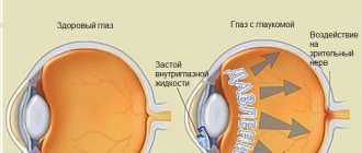
A disease in which the level of intraocular pressure is abnormally increased, which negatively affects the health of the organ of vision. As a result, the death of retinal nerve cells and their processes occurs. Increased pressure is the result of impaired outflow of intraocular fluid.
Important features of the disease:
- All animal species are susceptible.
- It occurs at any age (but the older the pet, the more factors for the development of pathology).
- Always progresses over time (treatment may slow this process).
- Until a certain point, it is invisible in animals.
Stages of development of glaucoma:
- Initial.
- Physiological changes causing disruption of the outflow of intraocular fluid.
- Increased intraocular pressure leads to disruption of blood flow and intercellular metabolism.
- Dysfunction of the nerve endings of the retina, degeneration of the optic nerve and its atrophy.
- Narrowing of the visual field and blindness.
Its types
Based on the cause of development, glaucoma is divided into two main types.
► Primary. Occurs primarily when there is no other eye disease affecting the outflow of fluid. In most cases, this process is bilateral (affects both eyes) and has a breed predisposition.
- Primary open-angle glaucoma. Found in Basset Hounds, Cocker Spaniels, Boston Terriers, Beagles, Chow Chows, Siberian Huskies, and Poodles. It develops rapidly and leads to loss of vision.
- Primary with a narrow angle.
- Primary angle-closure glaucoma (Samoyed husky, Labrador, toy poodle, beagle, basset hound).
► Secondary. When another eye or systemic disease interferes with the flow of intraocular fluid. It can be present in one or both eyes. It is observed in the Akita, Alaskan Malamute, Welsh Corgi, Dalmatian, Jack Russell Terrier and others breeds. The disease manifests itself between the ages of 4 and 10 years.
Classification of glaucoma
Glaucoma is divided into several types: primary and secondary. Primary is characterized by the formation of high pressure, but the eye itself is healthy. However, certain breeds of dogs are more susceptible to the disease compared to others. The reasons are hereditary, as they contain certain anomalies in the structure of the iridocorneal angle, where excess fluid drains. So, at a narrow angle, the outflow of liquid becomes somewhat more difficult, which provokes an increase in pressure. Secondary glaucoma is caused by damage to the eye itself, which consequently led to the disease. This may include injury, illness, or damage.
The classification also includes another variety - according to the angle of the anterior chamber, which can be closed, open, or narrow. Moreover, both types of classification exist in parallel and occur in different forms.
Therefore, in the case of a hereditary predisposition to such a disease, you will have to especially carefully monitor the pet’s health. If the first signs are detected, examination and subsequent therapy will be required.
Primary open-angle glaucoma is considered a purely hereditary disease, which is more often observed in beagles and poodles. The disease is characterized by a chronic form, so the pressure increases gradually and takes a long time. Moreover, the dog is able to see even in the last stages of the disease.
Goniodysplasia is a variation of the form of the disease just described. It primarily affects Great Danes, Cocker Spaniels, Samoyeds, Beagles and Labradors. At the same time, it is typical for the disease to have a recurrent manifestation in the form of severe attacks. So the eye swells, the pupils tend to dilate due to a change in the angle of refraction of light, and hyperemia is noted. Sometimes it even leads to blindness in the pet.
Causes of pathology in dogs
Factors that can lead to the development of glaucoma are:
- Breed predisposition. A number of breeds have a higher risk of being diagnosed with glaucoma than others.
- During observations, it was found that females are more susceptible to the closed-angle type of pathology.
- Genetic predisposition. The disease can be inherited.
- Various pathologies of the organ of vision (uveitis, retinal detachment, cataracts, etc.).
- Injuries (directly to the organ of vision or in the head and neck area).
- Other systemic diseases (diabetes mellitus, chronic renal failure, cardiovascular diseases).
- Sometimes glaucoma can become a complication after suffering from piroplasmosis (a dog becomes infected with parasites that damage blood cells through a tick bite).
Types of drops for glaucoma
Eye drops for glaucoma are divided into two types according to their mechanism of action.
Drugs that reduce the production of intraocular secretions:
- non-selective and selective beta-blockers;
- alpha and beta adrenergic blockers;
- topical carbonic anhydrase inhibitors;
- selective sympathomimetics.
Drugs that increase the outflow of intraocular fluid include:
- prostaglandins;
- m-cholinomimetics.
There are also combination drugs. They contain in one bottle medicinal substances that belong to different pharmacological groups. The use of combined components enhances the ability of drugs to reduce intraocular pressure.
{banner_horizontalnyy}
Symptoms of glaucoma in dogs
The initial signs of the disease are difficult for owners to notice. Therefore, people seek help when the pathology is already developing. During this period they note:
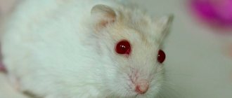
- Mydriasis is dilation of the pupil.
- The eye is enlarged. The body's defense mechanism reduces pain shock in this way.
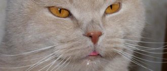
- Uveitis is redness of the eye.
- In severe and prolonged cases, the cornea gradually acquires a dull blue color.
- Vasodilation on the mucous membrane and whites of the eyeball.
- Depressed state of the animal. The pet refuses to play and is not active during walks.
- Pain in the area of the affected eye.
- Reduced vision or complete blindness.
Symptoms
With the acute development of glaucoma, the animal suddenly loses its vision. But this process can be reversed, while with chronic slow development, vision is lost gradually and irreversibly.
Regardless of the cause, the manifestation of glaucoma is always the same. You may not notice everything at once, but if you notice anything from this list, then hurry up and seek help from a veterinarian. Symptoms of increased intraorbital pressure:
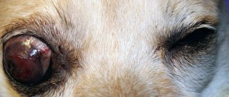
- Buphthalmos is an increase in size of the eyeball, this phenomenon is also called “bull’s eye.”
- Corneal edema.
- Redness.
- The vascular lines of the eye are visible.
- Soreness.
- Photophobia, the dog tries to hide in dark places.
- Mydriasis is dilation of the pupil.
Loss of vision. No matter how strange it may sound, you may not immediately notice that your pet’s vision has become much worse.
How can you test your pet's eyesight at home?
Glaucoma is not a disease that you can observe at home and expect that everything will go away on its own. At the first sign that your dog is seeing poorly, you should immediately contact a veterinary clinic.
Symptoms of decreased or loss of vision are:
- Photophobia. The pet avoids bright sources of light, hides in dark places, and squints during daytime walks.
- Poor orientation in space with good daytime visibility. Bumps into large objects and moves awkwardly. runs past the toy and does not immediately find a bowl of food or water.
- Glaucoma causes severe pain. This is indicated by a decrease in the pet’s activity, depressed mood, and decreased appetite.
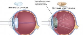
- When examining the eye, you may notice clouding of the pupil and anterior chamber.
Glaucoma in dogs cannot be treated at home. The dog experiences great discomfort and it is possible to select the necessary medications only after a highly specialized examination.
Reviews from doctors
Ilya, ophthalmologist: Many of my patients suffer from glaucoma. This is a disease in which intraocular pressure increases. This is dangerous by reducing visual acuity up to complete blindness. If the possibility of fluid outflow inside the eye is preserved, I prescribe medicinal drops. I often recommend Timolol, Trusopt. These are the most effective drugs, as they help not only reduce the production of intraocular fluid, but also its outflow from the eyes.
Olga, ophthalmologist: Patients with glaucoma often come to me for consultation and want to undergo surgery to restore the outflow of ocular fluid. In some cases, I do not recommend carrying out the procedure, since conservative treatment in the form of drops can be used. Then I advise you to use Pilocarpine, Timolol, Aceclidine. They effectively reduce intraocular pressure when used correctly.
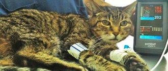
Diagnosis of pathology in a clinical setting
First of all, the doctor conducts a general examination of the animal. If the dog has previously had any eye problems, this should be noted. At the discretion of the veterinarian, general clinical and biochemical blood tests are taken.
Special examination methods include:
- Tonometry. The most important and informative way to diagnose glaucoma. Pressure measurements are taken in both eyes. The normal range is 10-25 mmHg. Art. If the value increases above 25, you can suspect a pathology in your pet. When performing tonometry, it is important to follow some rules. Fix the head carefully so that the pressure does not increase from excessive stress. It is advisable not to touch or turn the neck. Ideally, the dog's head is placed in a straight position, slightly held by the muzzle. It is also advisable not to touch the area around the eyes. The procedure for measuring intraocular pressure is not painful, animals tolerate it calmly. In some cases, anesthesia may be used.
- Gonioscopy. A study that allows you to check the angle of the anterior chamber of the eyeball. A special lens is placed on the eye and the structures are examined with a slit microscope.
- Ophthalmoscopy. A method by which a doctor will evaluate the condition of the fundus and retina.
- Ultrasound examination of the eyeball.
Diagnosis of glaucoma
The ophthalmology department of our clinic has all the necessary diagnostic equipment that allows you to conduct a complete ophthalmological examination and make a diagnosis of glaucoma.
A comprehensive examination usually includes the following main items:
- intraocular pressure examination;
- fundus examination;
- biomicroscopy of the anterior part of the eye;
- assessment of the condition of the optic nerve head
Glaucoma is a disease that can accompany an animal for the rest of its life. However, timely treatment and constant qualified observation can preserve vision and ensure an adequate quality of life for animals with glaucoma.
Treatment of glaucoma in dogs
If it is confirmed that a dog has glaucoma, therapy will be aimed at:
- decreased production of intraocular fluid;
- increasing its outflow;
- improvement and stabilization of eye hemodynamics;
- decrease in intraocular pressure;
- neuroprotection – protection of nerve cells from the effects of glaucomatous damage.
With such a pathology, it is important to start treatment as soon as possible.
► Medication method.
The use of special medications for glaucoma is advisable if the dog still has vision. Medicines, course duration and application features are selected individually.
Example of prescribed funds:
- Latanoprost.
- Mannitol.
- Pilocarpine.
- Metazopamid.
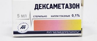
- Dexamethasone (if glaucoma is accompanied by an inflammatory process).
During the entire course of treatment, it is important to periodically monitor intraocular pressure until the condition stabilizes.
In case of a secondary type of disease, in addition to glaucoma, treatment is aimed at eliminating the root cause (uveitis, neoplasia, lens pathology, retinal detachment).
► Surgical method.
Indications for surgical therapy:
- If the use of local medications does not help and does not give positive dynamics.
- If the disease is too advanced and there is complete loss of vision in the affected eye. At this stage the process is irreversible.
- During the examination, neoplasia (neoplasms of any nature) was discovered.
Some surgical techniques used:
- Cyclocryosurgery involves freezing the ciliary body. As a result, the amount of intraocular fluid produced is reduced.
- Endolaser cyclophotocoagulation – destruction of the processes of the ciliary body. This procedure is the least invasive, but requires expensive equipment and excellent microsurgery skills from a specialist.
- Installation of the Ahmed valve. The design is installed on the eyeball and regulates the outflow of fluid. When too much of it accumulates, the valve helps remove the excess. The disadvantage of this method is that in animals the body does not always accept such foreign material. In some cases, inflammation may develop; as a result of this process, fibrin clogs the valve duct and it ceases to function. Also, this method is not widely used due to its high cost.
- Enucleation is the surgical removal of the eyeball with part of the eyelids, conjunctiva and Tenon's capsule. It is performed on a blind damaged eye in the terminal stage of glaucoma, when other treatment methods do not give a positive result or the increase in intraocular pressure is due to the presence of neoplasms. After surgical removal, the eyelids are completely sutured. The pet's eye looks like it is closed.
- If the owner wishes, after enucleation it is possible to perform prosthetics. In this case, a silicone intraocular prosthesis is installed. This procedure is carried out only for aesthetic purposes and has a number of nuances and contraindications.
Expert opinion
Kuzmenko Olga Olegovna
Information about the expert
Ask a Question
With any treatment method, it is imperative to monitor the condition of the second eye. Before surgery, the affected organ is stabilized as much as possible and pain is relieved.
The price of surgery for glaucoma in a dog is quite high and not affordable for everyone. The cost is influenced by many factors: equipment, qualifications of the veterinary ophthalmologist, the reputation of the clinic, the condition in which the patient is admitted. The most affordable option financially is enucleation. This operation is performed in almost every veterinary center and does not require a narrow specialty from a doctor.
Useful video on the topic: what is glaucoma in dogs, its diagnosis and treatment
Prevention
In order to minimize the risk of a dog or cat contracting glaucoma, animals should be treated for ophthalmological diseases in a timely manner and avoid situations where they could suffer eye injury. To improve the health of the visual organ, it is recommended to introduce special supplements into the diet of pets containing vitamins E and C, provitamin A (β-carotene), lutein, which increases visual acuity, and the microcirculation corrector rutin.
Glaucoma quickly leads to irreversible blindness due to optic nerve atrophy that develops against the background of high intraocular pressure. Vision lost as a result of this disease cannot be restored. Therefore, veterinarians recommend that everyone, especially older animals and animals genetically predisposed to glaucoma, measure intraocular pressure at least once a year.
Caring for a sick animal
- The owner of a sick dog must remember that this pathology is lifelong. Primary glaucoma in dogs can only be controlled with special eye drops, but in most cases vision will be lost over time.
- Prescribed drugs must be used as prescribed, since intraocular pressure increases very quickly, which will lead to blindness more quickly.
- Monitor the condition of the eye. If redness, an increase in the size of the eyeball, or clouding of the cornea are detected, you should immediately consult your doctor.
- Pet monitoring. The scheme of periodic examination and monitoring of intraocular pressure is established individually, depending on the severity of the process.
There is no prevention for primary glaucoma. The unaffected eye should be treated with antiglaucoma drugs (for example, dimercaine bromide - 1 drop every 24 hours, continuously). This will help delay the onset of glaucoma in a still healthy organ.
To avoid undesirable consequences in offspring, affected dogs are excluded from breeding.
Glaucoma in dogs is a pathology that leads to vision loss. The disease can be contained, but it is impossible to restore the main function of the affected organ. When you detect the first symptoms of the disease in your pet, it is important to seek help from a veterinarian as soon as possible, then the chances of saving the eye increase.
How to choose
Medicines should not be chosen by the patient, but only by the doctor. The choice is based on diagnostic studies obtained using laboratory and instrumental tests. The doctor takes into account the following patient indicators:
- age, some drops cannot be used for minors or elderly people;
- a health condition in which it is inadmissible to use certain types of drops that worsen the health condition (neuralgia, cardiovascular disorders, kidney and liver diseases);
- cost, many patients are over 60 years of age, so financial capabilities do not always allow them to purchase the most expensive drugs;
- the reason for the increase in intraocular pressure, why the doctor chooses the need to use a gland blocker or a means for the outflow of intraocular fluid;
- if the patient purchased the drug, but its effect gradually decreased, you can select another drug only together with the doctor (the reason may be the progression of glaucoma or addiction to the medicine).
If the drug is selected according to the patient's health condition, the possibility of side effects is significantly reduced. Despite this, the patient should periodically come for a follow-up examination with a doctor to identify a trend toward recovery.
Effective eye drops for glaucoma
There are a large number of effective eye drops for glaucoma. Compliance with the dosage regimen and doctor’s prescriptions helps normalize intraocular pressure and protect against blindness.
Azarga
Azarga eye drops contain brinzolamide, timolol maleate. This is a combination remedy: active components reduce intraocular pressure in various ways.
Used to reduce elevated intraocular pressure in open-angle glaucoma for patients in whom single-drug therapy has shown ineffectiveness.
Azarga is instilled one drop 2 times a day into the conjunctival sac. If you miss a dose, you should continue taking the drug as usual. Do not use if you are hypersensitive to the components or during pregnancy.
Azopt
The drug Azopt contains brynchzolamide in the form of a 1% solution. Refers to local inhibitors of carbonic anhydrase, an enzyme present in body tissues. Inhibition of the enzyme in the ciliary body of the eye contributes to a significant reduction in the amount of fluid. Thus, intraocular pressure decreases.
The drug should be instilled one drop 2 times a day. The bottle must be shaken before instilling the drug.
The drug should not be used during pregnancy or hypersensitivity to the component. The effectiveness of treatment in children has not been proven.
Betaxolol
Betaxolol is a beta1-blocker without intrinsic sympathomimetic activity.
Reduces high intraocular pressure. Used for open-angle glaucoma, and also as a component of combination therapy for conditions after laser trabeculoplasty. Use one drop 2 times a day. During the first month, treatment is carried out with constant monitoring of intraocular pressure. Further, the frequency of use is determined individually. The drug is prohibited for:
- decompensated heart failure;
- severe bradycardia;
- Prinzmetal's angina;
- severe forms of Raynaud's syndrome;
- hypotension.
During pregnancy, use is indicated in cases where the benefit to the mother is higher than the potential risk to the baby.
Betoptik
Betoptik eye drops containing 0.5% betaxolol. Indications, dosage and contraindications are similar to Betaxolol.
If clinical effectiveness is insufficient, additional therapy is prescribed.
The drug should be prescribed with caution in case of sinus bradycardia, AV block II and III degrees, severe heart failure, cardiogenic shock, myasthenia gravis, and diabetes mellitus.
Dorzopt
Dorzopt eye drops contain 2% of the active substance dorzolamide.
They are classified as local carbonic anhydrase inhibitors. The drug reduces the production of intraocular fluid without causing an accommodation spasm. The drug is prescribed for:
- open-angle glaucoma;
- pseudoexfoliative glaucoma;
- ophthalmic hypertension.
The solution should be instilled one drop into the conjunctival sac 3 times a day.
Drops are contraindicated for:
- chronic renal failure;
- childhood;
- pregnancy and breastfeeding;
- hypersensitivity to the active component;
- taking carbonic anhydrase inhibitors.
Drops should be prescribed for liver failure, diabetes, and pathologies of the cornea.
Irifrin
Irifrin is a 2.5% solution of phenylephrine hydrochloride.
Refers to sympathomimetics. Has alpha adrenergic activity. After instillation, the active substance causes pupil dilation. This effect occurs within 10-60 minutes after a single instillation and lasts for two hours. Applicable for:
- diagnostic pupil dilation;
- conducting a provocative test in patients with a narrow anterior chamber angle and suspected angle-closure glaucoma;
- glaucoma-cyclic crisis.
The dosage regimen for these drops for glaucoma is determined individually.
{banner_horizontalnyy2}
Kosopt
Cosopt is a combination drug that includes the active ingredients dorzolamide and timolol. Refers to antiglaucoma drugs for topical use (carbonic anhydrase inhibitor and beta-blocker).
The drug reduces the production of intraocular fluid. Timolol maleate slightly increases fluid outflow.
Prescribed one drop 2 times a day.
Xalacom
Xalacom is a combination drug containing the active substances latanoprost and timolol. Refers to synthetic analogues of prostaglandin F2-alpha and beta adrenergic blockers. The mechanisms for reducing high intraocular pressure are different for the components included in the drops, which ensures a lasting therapeutic effect. The effect of the drops occurs within an hour after application and lasts up to eight hours. With repeated use, the effect lasts throughout the day.
Xalacom is prescribed to lower intraocular pressure in patients with open-angle glaucoma.
The drug should be instilled one drop once a day. Xalacom is contraindicated for:
- bronchial asthma;
- severe chronic obstructive pulmonary disease;
- sinus bradycardia;
- severe heart failure;
- cardiogenic shock;
- atrioventricular block 2 and 3 degrees;
- sick sinus syndrome.
Xalatan
Xalatan eye drops contain 0.005% latanoprost. This component is a selective FP (prostaglandin F) receptor agonist. It effectively lowers intraocular pressure as it enhances the process of outflow of aqueous humor. The pressure inside the eye decreases a few hours after instillation.
The drug should be instilled one drop once a day.
Drops are prohibited in case of hypersensitivity to the active component. Contraindications for use:
- age up to 1 year (efficacy and safety have not been established);
- hypersensitivity to latanoprost or other components of the drug.
Pilocarpine
Pilocarpine is an m-cholinomimetic drug.
Effectively improves the evacuation of intraocular fluid and reduces intraocular pressure. Improves trophism of eye tissue. Indicated for:
- primary and chronic open-angle glaucoma;
- secondary glaucoma;
- acute attack of angle-closure glaucoma;
- the need for pupil constriction.
Pilocarpine solution should be instilled into the eye, 1 to 2 drops, 2 to 4 times a day.
Scheme of use for an attack of angle-closure glaucoma:
- during the first hour - drop by drop after 15 minutes;
- for 2 – 3 hours – drop by drop every half hour;
- for 4 – 6 hours – a drop every hour;
- in the future - from 3 to 6 times a day until the attack stops.
Timolol
Timolol is a non-selective beta blocker.
Applicable for:
- chronic open-angle glaucoma;
- angle-closure glaucoma;
- secondary glaucoma.
The method of application depends on the patient's condition.
Travatan
The active substance is travoprost.
It is a prostaglandin FP receptor agonist. Indicated for open-angle glaucoma. Travatan is prohibited when:
- pregnancy and breastfeeding:
- increased individual sensitivity.
{banner_horizontalnyy3}
Fotil
Active substances: pilocarpine, timolol. It is used for open-angle, closed-angle or secondary glaucoma, as well as for increased intraocular pressure after eye surgery.
Fotil should be instilled drop by drop 2 times a day into the affected eye.
Contraindications for use:
- sinus bradycardia;
- AV block II and III degrees;
- decompensated heart failure;
- cardiogenic shock;
- bronchial asthma;
- COPD;
- children and adolescents under 18 years of age (due to the lack of data on the effectiveness and safety of the drug in children and adolescents);
- condition immediately after ophthalmological operations and other eye diseases in which constriction of the pupil is undesirable;
- hypersensitivity to the components of the drug.